Перейти к содержимому
КОСТИ НИЖНЕЙ КОНЕЧНОСТИ
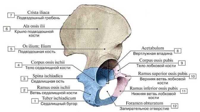 Тазовая кость, правая. Цветом выделены отдельные кости:
1 — Ischial tuberosity; 2— Ischium, ramus; 3— Ischial spine; 4 — Bodyofischium; 5 — Ilium; 6 — Ala ofilium; Wingofilium; 7— Iliae crest; 8 — Acetabulum; 9 — Pubis, body; 10— Superior pubic ramus; 11— Inferior pubic ramus; 12 — Obturator foramen
Тазовая кость, правая. Цветом выделены отдельные кости:
1 — Ischial tuberosity; 2— Ischium, ramus; 3— Ischial spine; 4 — Bodyofischium; 5 — Ilium; 6 — Ala ofilium; Wingofilium; 7— Iliae crest; 8 — Acetabulum; 9 — Pubis, body; 10— Superior pubic ramus; 11— Inferior pubic ramus; 12 — Obturator foramen
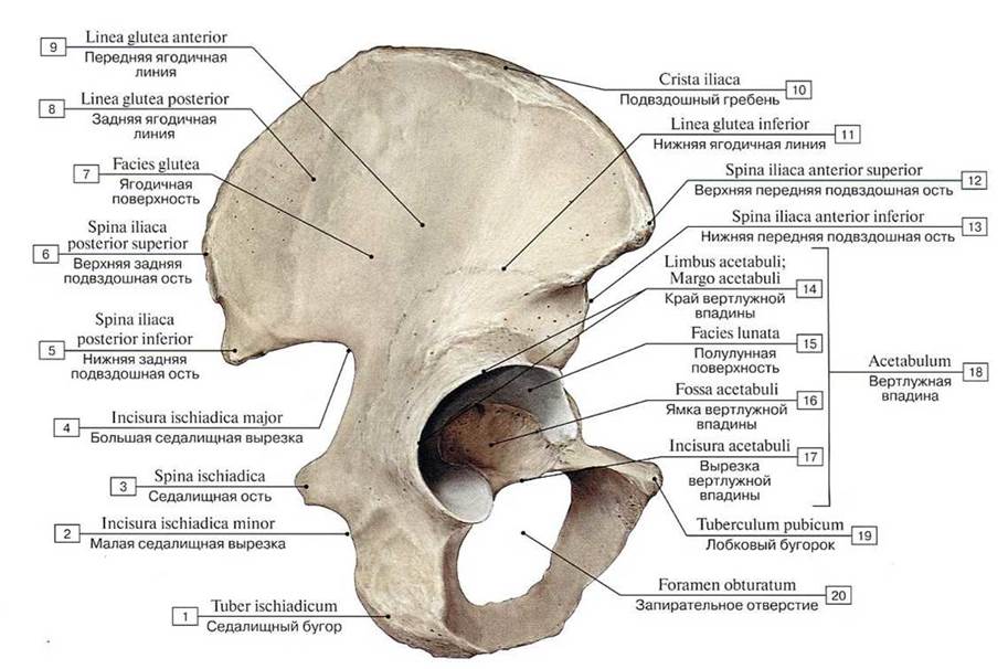 Тазовая кость, правая, вид с латеральной стороны:
1 — Ischial tuberosity; 2 — Lesser sciatic notch; 3 — Ischial spine; 4 — Greater sciatic notch; 5 — Posterior inferior iliac spine; 6 —- Posterior superior iliac spine: 7 — Gluteal surface; 8 — Posterior glutcal line; 9 — Anterior gluteal line; 10 — Iliac crest; 11 — Inferior gluteal line; 12 — Anterior superior iliac spine; 13 — Anterior inferior iliac spine; 14 — Acetabular margin; 15 — Lunate surface; 16 — Acetabular fossa; 17— Acetabular notch; 18= 14 + 15 + 16 + 17 — Acetabulum; 19 — Pubie tubercle; 20 — Obturator foramen
Тазовая кость, правая, вид с латеральной стороны:
1 — Ischial tuberosity; 2 — Lesser sciatic notch; 3 — Ischial spine; 4 — Greater sciatic notch; 5 — Posterior inferior iliac spine; 6 —- Posterior superior iliac spine: 7 — Gluteal surface; 8 — Posterior glutcal line; 9 — Anterior gluteal line; 10 — Iliac crest; 11 — Inferior gluteal line; 12 — Anterior superior iliac spine; 13 — Anterior inferior iliac spine; 14 — Acetabular margin; 15 — Lunate surface; 16 — Acetabular fossa; 17— Acetabular notch; 18= 14 + 15 + 16 + 17 — Acetabulum; 19 — Pubie tubercle; 20 — Obturator foramen
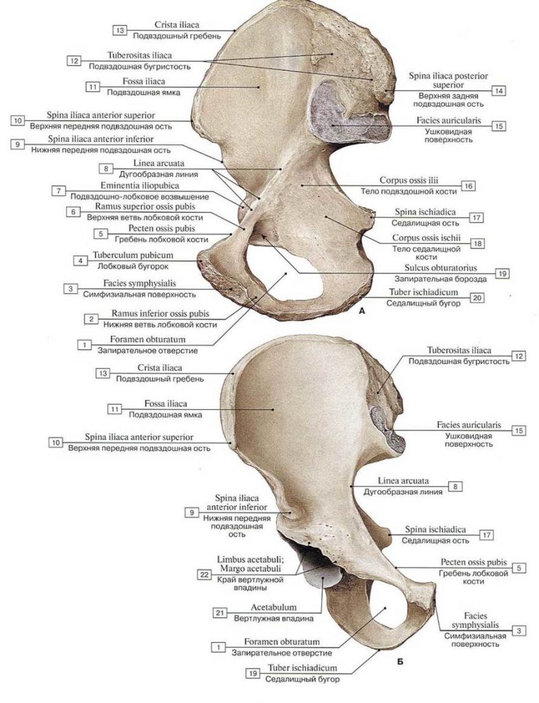 Тазовая кость, правая (А — вид с медиальной стороны, Б — вид спереди):
1 — Obturator foramen; 2 — Inferior pu- bic ramus; 3 — Symphysial surface; 4 — Pu- bic tubercle; 5 — Pecten pubis; Pectinea! line; 6 — Superior pubic ramus; 7— Iliopubiceminen- ce; 8 — Arcuate line; 9 — Anterior inferior iliac spine; 10 — Anterior superior iliac spine; 11 — Iliac fossa; 12 — Iliac tubcrosity; 13 — Iliac crest; 14 —- Posterior superior iliac spine; 15 — Ilium, auricular surface; 16— Bodyof ilium; 17 — Ischial spine; 18 — Bodyof ischium; 19 — Obturator groove; 20 — Ischial tuber; 21 — Acetabulum;
Тазовая кость, правая (А — вид с медиальной стороны, Б — вид спереди):
1 — Obturator foramen; 2 — Inferior pu- bic ramus; 3 — Symphysial surface; 4 — Pu- bic tubercle; 5 — Pecten pubis; Pectinea! line; 6 — Superior pubic ramus; 7— Iliopubiceminen- ce; 8 — Arcuate line; 9 — Anterior inferior iliac spine; 10 — Anterior superior iliac spine; 11 — Iliac fossa; 12 — Iliac tubcrosity; 13 — Iliac crest; 14 —- Posterior superior iliac spine; 15 — Ilium, auricular surface; 16— Bodyof ilium; 17 — Ischial spine; 18 — Bodyof ischium; 19 — Obturator groove; 20 — Ischial tuber; 21 — Acetabulum;
Кости свободной части нижней конечности
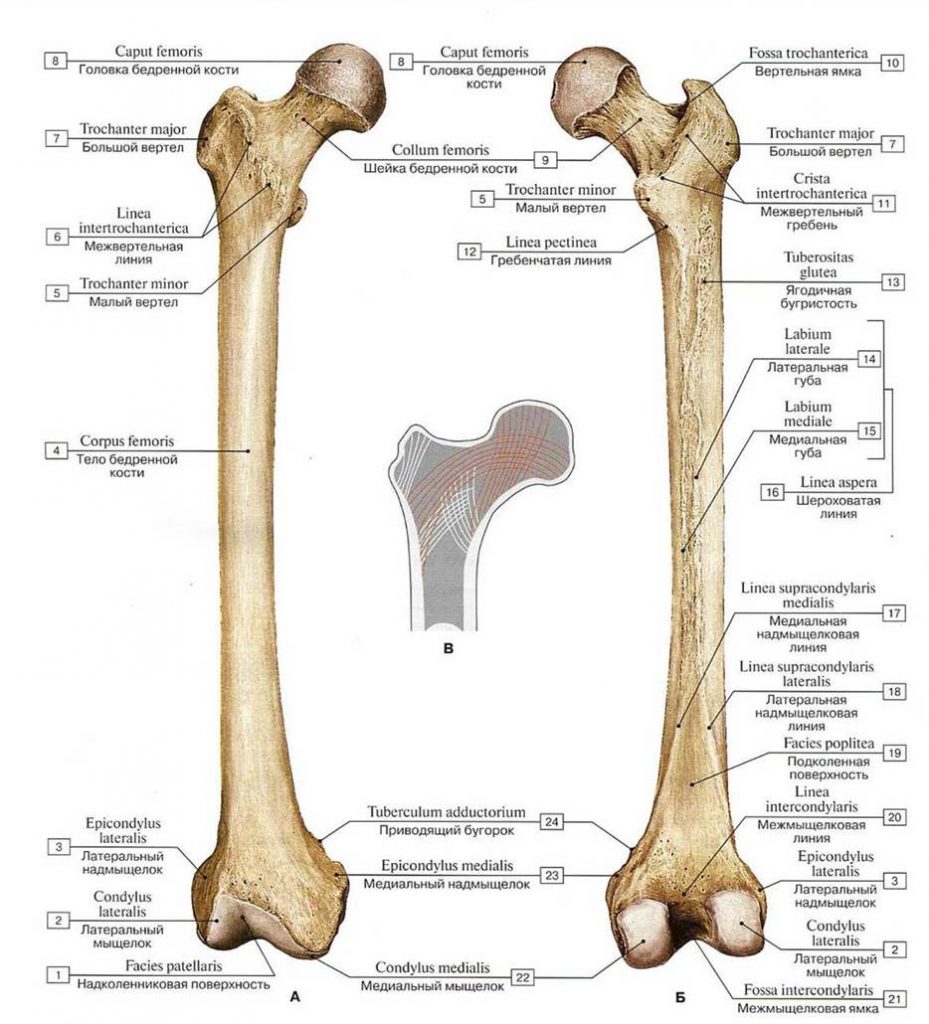 Бедренная кость, правая (А — вид спереди, Б — вид сзади, В — направление костных трабекул головки и шейки бедренной кости относительно прилагаемой нагрузки):
1 — Patellar surface; 2 — Lateral condyle; 3 — Lateral epicondyle; 4 — Shaft of femur; Body of femur; 5 — Lesser trochanter; 6 — lnter- trochanteric line; 7 — Greatcr trochanter; 8 — Head of femur; 9 — Neck of femur; 10 — Trochanteric fossa; 11 — Intertrochanteric crest; 12— Pectineal line; Spiral line; 13 — Gluteal tuberosity; 14 — Lateral lip; 15 — Medial lip; 16 —Linea aspera; 17 — Medial supracondylar line; 18 — Lateral supracondylar line; 19 — Poplileal surface; 20 — Intercondylar line; 21 — Intercondylar fossa; 22 — Medial condyle; 23 — Medial epicondyle; 24 — Adductor tubercle
Бедренная кость, правая (А — вид спереди, Б — вид сзади, В — направление костных трабекул головки и шейки бедренной кости относительно прилагаемой нагрузки):
1 — Patellar surface; 2 — Lateral condyle; 3 — Lateral epicondyle; 4 — Shaft of femur; Body of femur; 5 — Lesser trochanter; 6 — lnter- trochanteric line; 7 — Greatcr trochanter; 8 — Head of femur; 9 — Neck of femur; 10 — Trochanteric fossa; 11 — Intertrochanteric crest; 12— Pectineal line; Spiral line; 13 — Gluteal tuberosity; 14 — Lateral lip; 15 — Medial lip; 16 —Linea aspera; 17 — Medial supracondylar line; 18 — Lateral supracondylar line; 19 — Poplileal surface; 20 — Intercondylar line; 21 — Intercondylar fossa; 22 — Medial condyle; 23 — Medial epicondyle; 24 — Adductor tubercle
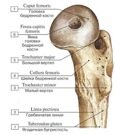 Верхний эпифиз бедренной кости, правой, вид с медиальной стороны:
1 — Gluteal tuberosity; 2 — Pectineal line; Spiral line; 3 — Lesser trochanter; 4 — Neck of femur; 5 — Greater trochanter; 6 — Fovea forligament ofhead; 7— Headoffemur
Верхний эпифиз бедренной кости, правой, вид с медиальной стороны:
1 — Gluteal tuberosity; 2 — Pectineal line; Spiral line; 3 — Lesser trochanter; 4 — Neck of femur; 5 — Greater trochanter; 6 — Fovea forligament ofhead; 7— Headoffemur
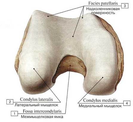 Нижний эпифиз бедренной кости, правой, вид спереди:
1 — lntcrcondylar fossa; 2 — Lateral condyle; 3 — Patellar suiface; 4 — Mcdial condyle
Нижний эпифиз бедренной кости, правой, вид спереди:
1 — lntcrcondylar fossa; 2 — Lateral condyle; 3 — Patellar suiface; 4 — Mcdial condyle
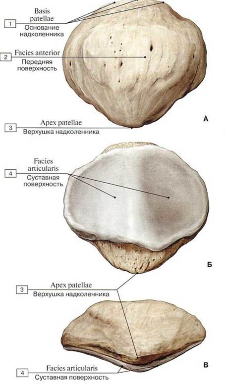 Надколенник, правый (А — передняя поверхность, Б — суставная поверхность, В — вид сбоку)
1 — Base of patella; 2 — Anterior surface; 3 — Apex of patella; 4 — Articular surface
Надколенник, правый (А — передняя поверхность, Б — суставная поверхность, В — вид сбоку)
1 — Base of patella; 2 — Anterior surface; 3 — Apex of patella; 4 — Articular surface
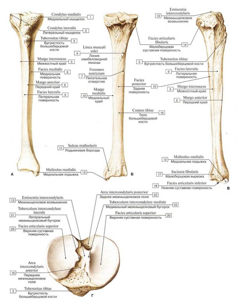 Большеберцовая кость, правая (А — вид спереди, Б — вид сзади, В — вид с латеральной стороны, Г — проксимальный эпифиз, вид сверху):
1 — Medial condyle; 2 — Lateral condyle; 3 — Tibial tuberosity; 4 — Soleal line; 5 — Interosseous bordcr; 6 — Medial surfacc: 7 — Nutrient foramen; 8 — Anterior border; 9 — Lateral surface; 10 — Medial border; 11— Malleolar groove; 12 — Medial malleolus; 13— Intercondylareminence; 14 — Fibular articular facet; 15 — Posterior surface; 16 — Shaft of tibia; Body of tibia; 17— Fibular notch; 18 — Inferior articular surface; 19 — Anterior intercondylar area; 20 — Superior articular surface; 21 — Lateral intercondylar tubercle; 22 — Lateral intercondylar area; 23 — Medial intercondylar tubercle
Большеберцовая кость, правая (А — вид спереди, Б — вид сзади, В — вид с латеральной стороны, Г — проксимальный эпифиз, вид сверху):
1 — Medial condyle; 2 — Lateral condyle; 3 — Tibial tuberosity; 4 — Soleal line; 5 — Interosseous bordcr; 6 — Medial surfacc: 7 — Nutrient foramen; 8 — Anterior border; 9 — Lateral surface; 10 — Medial border; 11— Malleolar groove; 12 — Medial malleolus; 13— Intercondylareminence; 14 — Fibular articular facet; 15 — Posterior surface; 16 — Shaft of tibia; Body of tibia; 17— Fibular notch; 18 — Inferior articular surface; 19 — Anterior intercondylar area; 20 — Superior articular surface; 21 — Lateral intercondylar tubercle; 22 — Lateral intercondylar area; 23 — Medial intercondylar tubercle
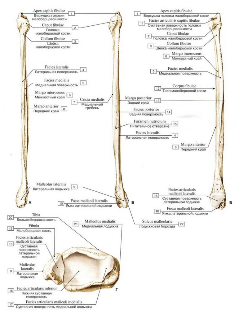 Малоберцовая кость, правая (А — вид спереди, Б — вид сзади, В — вид с медиальной стороны,
Г — суставные поверхности нижних эпифизов костей голени):
1 — Apex ofhead; 2 — Head of fibula; 3 — Neck of fibula; 4 — Lateral surface; 5 — Medial surface; 6 — Interosseous bordcr; 7— Medial crcst; 8 — Anterior border; 9 — Lateral malleolus; 10— Malleolar fossa; 11 — Articular facet; 12 — Shaft of fibula; Body of fibula; 13 — Posterior border; 14 — Posterior surface; 15 — Nutrient foramen; 16 — Articular facet; 17 — Articular facet; 18 — Inferior articular surface; 19 — Fibula; 20 — Tibia; 21 — Medial malleolus; 22— Malleolar groove
Малоберцовая кость, правая (А — вид спереди, Б — вид сзади, В — вид с медиальной стороны,
Г — суставные поверхности нижних эпифизов костей голени):
1 — Apex ofhead; 2 — Head of fibula; 3 — Neck of fibula; 4 — Lateral surface; 5 — Medial surface; 6 — Interosseous bordcr; 7— Medial crcst; 8 — Anterior border; 9 — Lateral malleolus; 10— Malleolar fossa; 11 — Articular facet; 12 — Shaft of fibula; Body of fibula; 13 — Posterior border; 14 — Posterior surface; 15 — Nutrient foramen; 16 — Articular facet; 17 — Articular facet; 18 — Inferior articular surface; 19 — Fibula; 20 — Tibia; 21 — Medial malleolus; 22— Malleolar groove
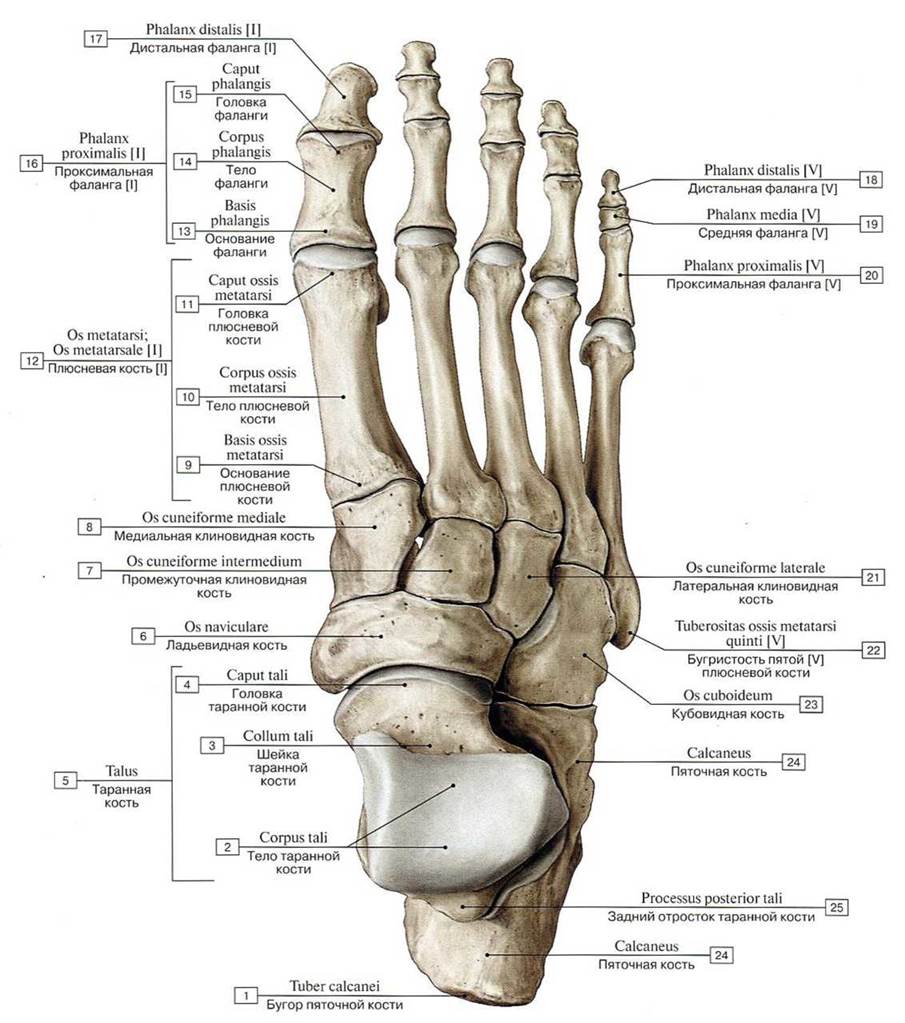 Кости стопы, правой, вид сверху:
1 — Calcaneal tuber; 2 — Body оf talus; 3 — Neck of talus; 4 — Head of talus; 5 — Talus; 6 — Navicular; 7 — Intermediate cuneiform; Middle cuneiform; 8 — Medial cuneiform; 9 — Base of metatarsal; 10 — Shaft of metatarsal; Body of metatarsal; 11 — Head of metatarsal; 12 — Metatarsal [1]; 13 — Base of phalanx; 14 — Shaft of phalanx; Body of phalanx; 15 — Head of phalanx; 16 — Proximal phalanx[1]; 17 — Distal phalanx [l]; 18 — Distal phalanx [V]; 19 — Middle phalanx[V]; 20— Proximal phalanx [V]; 21 — Lateral cuneiform; 22— Tubero-sity of fifth metatarsal bone [V]; 23 — Cuboid; 24 — Calcaneus; 25— Posterior process
Кости стопы, правой, вид сверху:
1 — Calcaneal tuber; 2 — Body оf talus; 3 — Neck of talus; 4 — Head of talus; 5 — Talus; 6 — Navicular; 7 — Intermediate cuneiform; Middle cuneiform; 8 — Medial cuneiform; 9 — Base of metatarsal; 10 — Shaft of metatarsal; Body of metatarsal; 11 — Head of metatarsal; 12 — Metatarsal [1]; 13 — Base of phalanx; 14 — Shaft of phalanx; Body of phalanx; 15 — Head of phalanx; 16 — Proximal phalanx[1]; 17 — Distal phalanx [l]; 18 — Distal phalanx [V]; 19 — Middle phalanx[V]; 20— Proximal phalanx [V]; 21 — Lateral cuneiform; 22— Tubero-sity of fifth metatarsal bone [V]; 23 — Cuboid; 24 — Calcaneus; 25— Posterior process
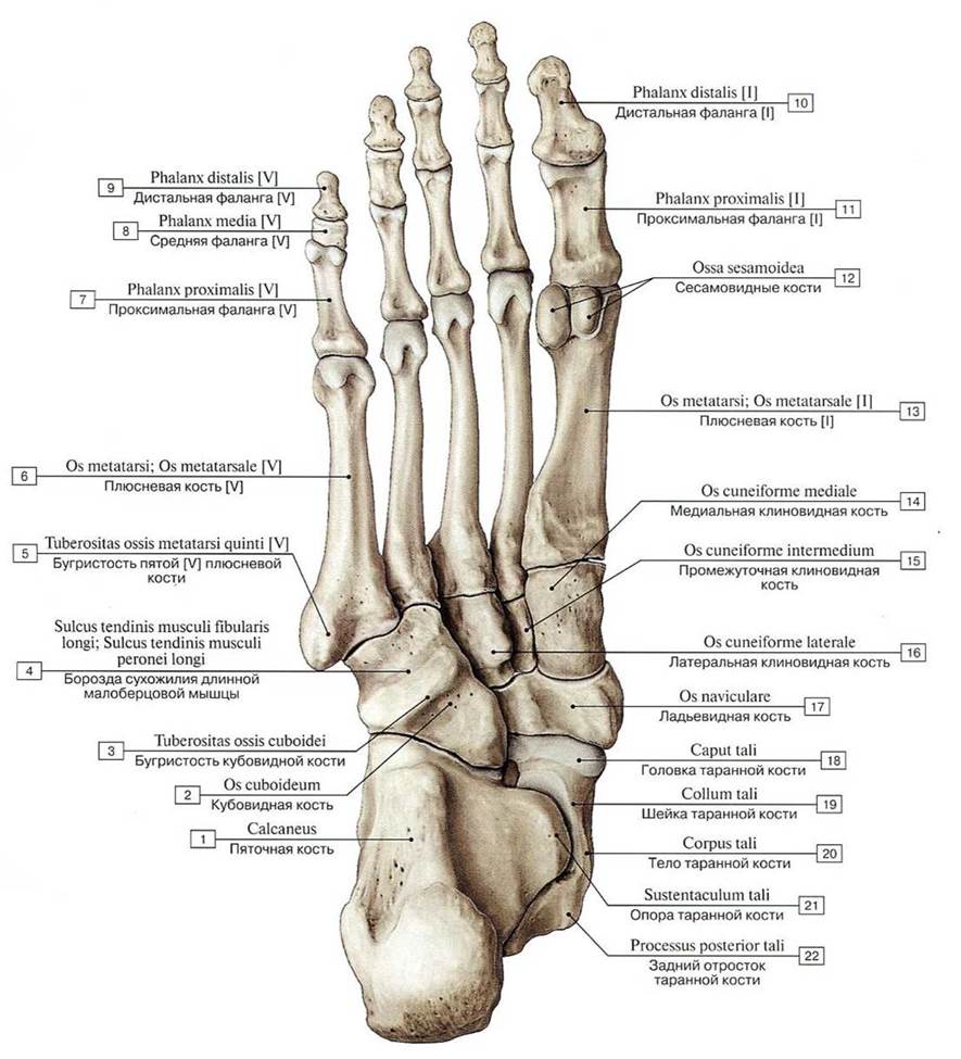 Кости стопы, правой, вид снизу:
1 — Calcaneus; 2 — Cuboid: 3 — Tuberosily of cuboid: 4 — Groovc for tendon of flbularis longus; Groove for tendon of peroneus longus; 5 — Tuberosity of fifth metatarsal bone [V]; 6 — Metatarsal [V]; 7 — Proximal phalanx [V]; 8 — Middle phalanx [V]; 9 — Distal phalanx [V]; 10 — Distal phalanx [I]; 11 — Proximal phalanx [I]; 12 — Sesamoid bones; 13 — Metatarsal [I]; 14— Medial cuneiform; 15 — Intermediate cuneiform; Middle cuneiform; 16 — Lateral cuneiform; 17— Navicular; 18 — Headoftalus; 19— Neckoftalus; 20— Bodyoftalus; 21 — Sustentaculum tali; Talar shclf; 22 —Talus, posterior process
Кости стопы, правой, вид снизу:
1 — Calcaneus; 2 — Cuboid: 3 — Tuberosily of cuboid: 4 — Groovc for tendon of flbularis longus; Groove for tendon of peroneus longus; 5 — Tuberosity of fifth metatarsal bone [V]; 6 — Metatarsal [V]; 7 — Proximal phalanx [V]; 8 — Middle phalanx [V]; 9 — Distal phalanx [V]; 10 — Distal phalanx [I]; 11 — Proximal phalanx [I]; 12 — Sesamoid bones; 13 — Metatarsal [I]; 14— Medial cuneiform; 15 — Intermediate cuneiform; Middle cuneiform; 16 — Lateral cuneiform; 17— Navicular; 18 — Headoftalus; 19— Neckoftalus; 20— Bodyoftalus; 21 — Sustentaculum tali; Talar shclf; 22 —Talus, posterior process
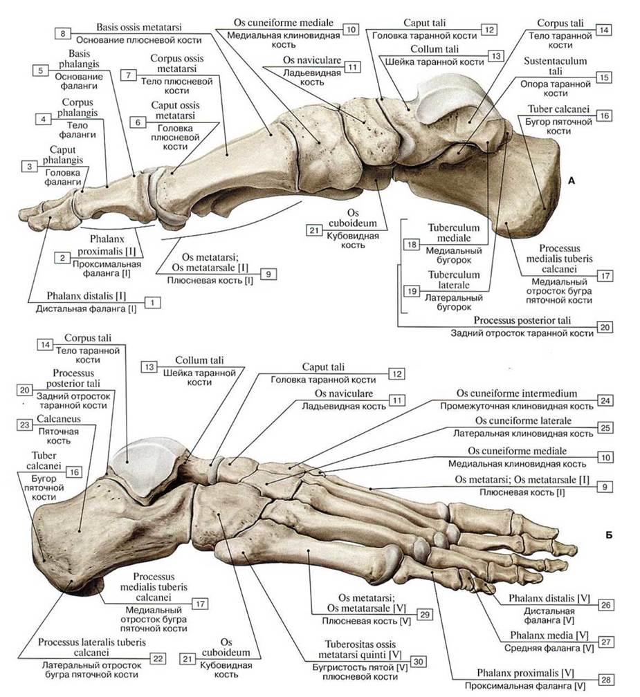 Кости стопы, правой (А — вид с медиальной стороны, Б — вид с латеральной стороны):
1 — Distal phalanx [I]; 2 — Proximal phalanx [I]; 3 — Head of phalanx; 4 — Shaft of phalanx; Body of phalanx; 5 — Base of phalanx; 6 — Head of metatarsal; 7 — Shaft of metatarsal; Body of metatarsal; 8 — Base of metatarsal; 9 — Metatarsal [I]; 10 — Medial cunciform; 11 — Navicular; 12 — Head of talus; 13 — Neck of talus; 14 — Body of talus; 15 — Sustentaculum tali; Talar shelf; 16 — Calcaneal tuber; 17— Calcaneus, medial process; 18 — Medial tubercle; 19— Lateral tubercle; 20 — Talus, posterior process; 21 — Cuboid; 22— Calcaneus, lateral process; 23— Calcaneus; 24— Intermediate cuneiform; Middle cuneiform; 25 — Lateral cuneiform; 26 — Distal phalaax [V]; 27 — Middle phalanx [V]; 28 — Proximal phalanx [V]; 29— Metatarsal [V]; 30— Tuberosity offifth metatarsal bone [V]
Кости стопы, правой (А — вид с медиальной стороны, Б — вид с латеральной стороны):
1 — Distal phalanx [I]; 2 — Proximal phalanx [I]; 3 — Head of phalanx; 4 — Shaft of phalanx; Body of phalanx; 5 — Base of phalanx; 6 — Head of metatarsal; 7 — Shaft of metatarsal; Body of metatarsal; 8 — Base of metatarsal; 9 — Metatarsal [I]; 10 — Medial cunciform; 11 — Navicular; 12 — Head of talus; 13 — Neck of talus; 14 — Body of talus; 15 — Sustentaculum tali; Talar shelf; 16 — Calcaneal tuber; 17— Calcaneus, medial process; 18 — Medial tubercle; 19— Lateral tubercle; 20 — Talus, posterior process; 21 — Cuboid; 22— Calcaneus, lateral process; 23— Calcaneus; 24— Intermediate cuneiform; Middle cuneiform; 25 — Lateral cuneiform; 26 — Distal phalaax [V]; 27 — Middle phalanx [V]; 28 — Proximal phalanx [V]; 29— Metatarsal [V]; 30— Tuberosity offifth metatarsal bone [V]
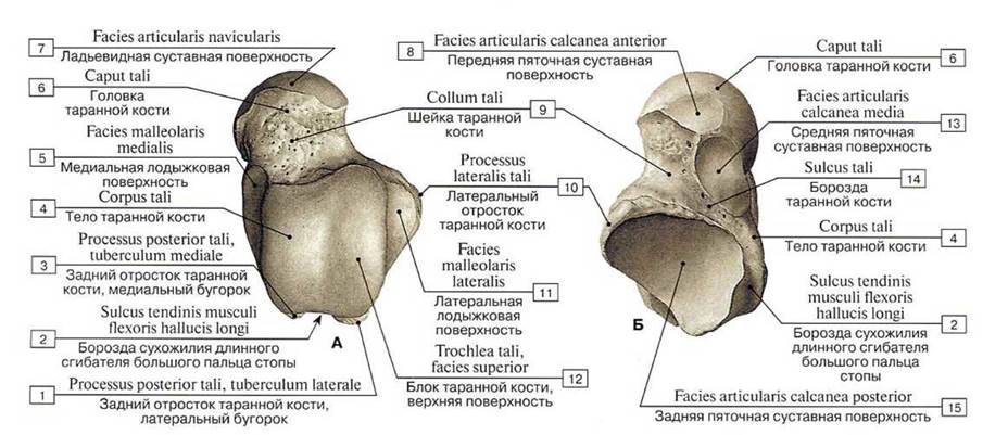 Таранная кость, правая (А — вид сверху, Б — вид снизу):
1 — Posterior process, lateral tubercle; 2 — Groove for tendon of flexor hallucis longus; 3 — Posterior process, medial tubercle; 4 — Body of talus; 5 — Medial malleolar facet; 6 — Head of talus; 7 — Navicular articular surface; 8 — Anterior lacet for calcaneus; 9 — Neck of talus; 10 — Lateral process; 11 — Lateral malleolar facet; 12 — Trochlea of talus, superior facet; 13 — Middle facet for calcaneus; 14 — Sulcus tali; 15 — Posterior calcaneal articular facet
Таранная кость, правая (А — вид сверху, Б — вид снизу):
1 — Posterior process, lateral tubercle; 2 — Groove for tendon of flexor hallucis longus; 3 — Posterior process, medial tubercle; 4 — Body of talus; 5 — Medial malleolar facet; 6 — Head of talus; 7 — Navicular articular surface; 8 — Anterior lacet for calcaneus; 9 — Neck of talus; 10 — Lateral process; 11 — Lateral malleolar facet; 12 — Trochlea of talus, superior facet; 13 — Middle facet for calcaneus; 14 — Sulcus tali; 15 — Posterior calcaneal articular facet
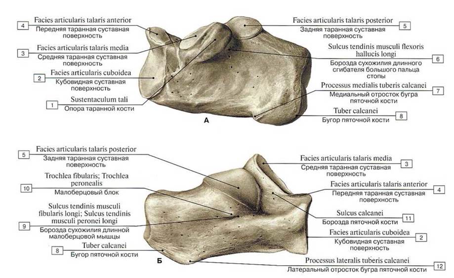 Пяточная кость, правая (А — вид с медиальной стороны, Б — вид с латеральной стороны):
1— Sustentaculum tali; Talar shelf; 2 — Articular surface for cuboid; 3 — Middle talar articular surfacc; 4 — Anterior talar articular surface; 5— Posterior talar articular surface; 6— Groove fortendon of flexor hallucis longus; 7— Medial process; 8 — Calcaneal tuber; 9 — Groove for tendon of fibularis longus; Groove fortendon of peroneus longus; 10 — Fibular trochlea; Peroneal trochlea; Peroneal tubercle; 11 — Calcaneal sulcus; 12 — Lateral process
Пяточная кость, правая (А — вид с медиальной стороны, Б — вид с латеральной стороны):
1— Sustentaculum tali; Talar shelf; 2 — Articular surface for cuboid; 3 — Middle talar articular surfacc; 4 — Anterior talar articular surface; 5— Posterior talar articular surface; 6— Groove fortendon of flexor hallucis longus; 7— Medial process; 8 — Calcaneal tuber; 9 — Groove for tendon of fibularis longus; Groove fortendon of peroneus longus; 10 — Fibular trochlea; Peroneal trochlea; Peroneal tubercle; 11 — Calcaneal sulcus; 12 — Lateral process
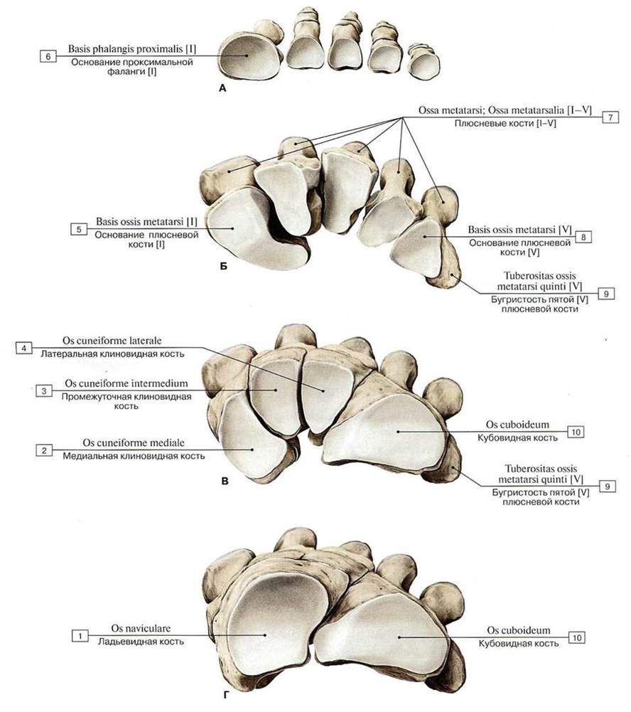 Кости стопы, правой, вид сверху (А — основания проксимальных фаланг, Б — основания костей плюсны,
В — клиновидные и кубовидные кости, Г — ладьевидная и кубовидная кости):
1 — Navicular; 2 — Medial cuneiform; 3 — Intermediate cuneiform; Middlc cuneiform; 4 — Lateral cuneiform; 5— Base of metatarsal [1]; 6 — Base of proximal phalanx [I]; 7 — Mctatarsals [I—V]; 8 — Base of metatarsal [V]; 9 — Tubcrosity of fifth metatarsal bone [V]; 10— Cuboid
Кости стопы, правой, вид сверху (А — основания проксимальных фаланг, Б — основания костей плюсны,
В — клиновидные и кубовидные кости, Г — ладьевидная и кубовидная кости):
1 — Navicular; 2 — Medial cuneiform; 3 — Intermediate cuneiform; Middlc cuneiform; 4 — Lateral cuneiform; 5— Base of metatarsal [1]; 6 — Base of proximal phalanx [I]; 7 — Mctatarsals [I—V]; 8 — Base of metatarsal [V]; 9 — Tubcrosity of fifth metatarsal bone [V]; 10— Cuboid