Атлас МИОЛОГИЯ: Г.Л. Билич – 2014

Мышцы задней стенки брюшной полости

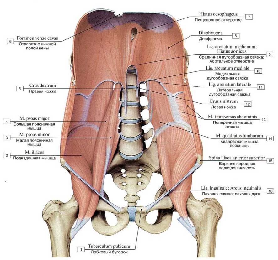
Мышцы задней стенки живота, вид спереди: 
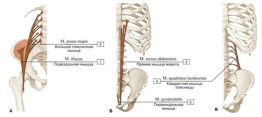
1 — Pubic tubercle; 2— Iliacus; 3 — Psoas minor; 4 — Psoas major; 5 — Right crus; 6— Caval opening; 7— Oesophageal hiatus; 8 — Diaphragm; 9 — Median arcuate ligament; Aortic hiatus; 10— Medial arcuate ligament; 11 — Lateral arcuate ligament; 12— Left crus; 13 — Transversus abdominis; Transverse abdominal; 14 — Quadratus lumborum; 15 — Anterior superior iliac spine; 16 — Inguinal ligament
Места начала и прикрепления (А — подвздошно-поясничной мышцы, Б — прямой мышцы живота и пирамидальной мышцы, Б — квадратной мышцы поясницы) (схемы): 
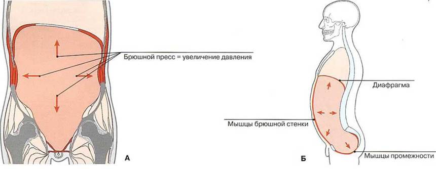
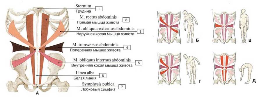
1 — Iliacus; 2 —- Psoas major; 3 — Rectus abdominis; 4 — Quadratus lumborum; 5 — Pyramidalis
Функции мышц брюшного пресса (А – фронтальная плоскость, Б – сагиттальная плоскость) (схема)

Движения туловища осуществляют косые и прямые мышцы живота
А. Направление пучков и организация прямых и косых мышц живота.
Б. Туловище наклонено вправо с одновременным поворотом влево благодаря сокращению наружной косой мышцы живота в правую сторону и внутренней косой мышцы живота в левую сторону.

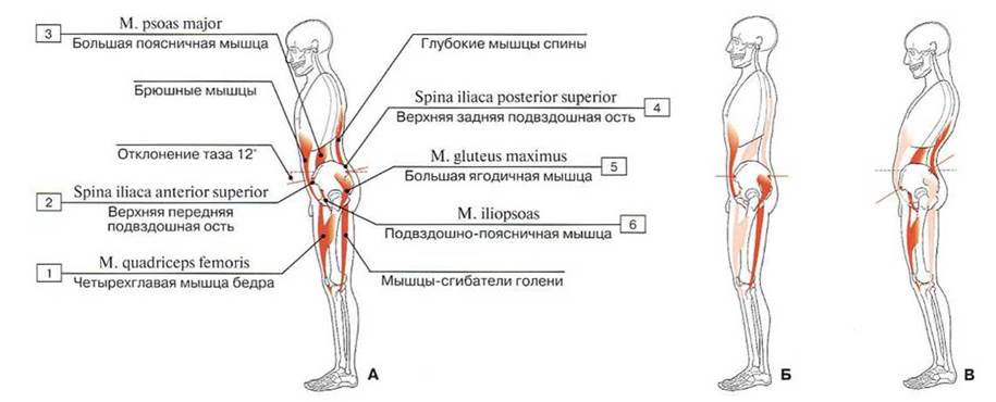
Участие мышц брюшной стенки в движениях таза: активные и пассивные положения (А — нормальное активное положение, Б — активное стабильное положение, В — пассивное «упавшее» положение) (схемы):1 — Quadriceps femoris; 2 — Anterior superior iliac spine; 3 — Psoas major; 4 — Posterior superior iliac spine; 5 — Gluteus maximus; 6 — Iliopsoas
Участие мышц переднебоковой стенки живота в движениях туловища (схемы): 1 — Sternum; 2 — Rectus abdominis; 3 — External oblique; 4 — Transversus abdominis; Transverse abdominal; 5 — Internal oblique; 6 — Linea alba; 7— Pubic symphysis

В. Вращение туловища вправо выполняется мышцами правой боковой стенки живота и левыми глубокими мышцами спины.

Г. Туловище наклонено вправо благодаря сокращению правой боковой стенки живота (этому способствуют и правые глубокие мышцы спины).

Д. Сгибание туловища осуществляется благодаря билатеральному сокращению прямых мышц живота, мышц боковых стенок живота и подвздошных мышц.

Внутренняя поверхность передней брюшной стенки мужчины: 1 — Obturator branch; 2 — Obturator nerve, artery and veins; 3 — Ductus deferens; Vas deferens; 4 — Artery and vein to ductus deferens; 5 — Femoral artery and vein; 6 — Testicular artery and vein; 7— Deep circumflex iliac artery and vein; 8— Inferior epigastrie artery and vein; 9 — Rectus abdominis; 10 — Arcuate line; 11 — Rectus sheath; 12 — Transversalis fascia; 13 — Parietal peritoneum; 14 — Diaphragm; 15 — Falciform ligantent; 16 — Round ligament of the liver; Para-umbilical veins; 17 — Umbilical region; 18 — Median umbilical fold; 19 — Medial umbilical fold; 20 — Lateral umbilical fold; Epigastrie fold; 21 — Iliacus; 22 — Lateral inguinal fossa; Deep inguinal ring; 23 — Psoas major; 24— Medial inguinal fossa; 25 — Supravesical fossa; 26 — Urinary bladdcr; 27— Prostate
. Апоневрозы наружных косых мышц живота, белая линия живота, паховый канал женщины, вид спереди: 1 — Falciform margin, inferior hom; 2 — Fascia lata; 3 — Femoral artery; 4 — Femoral vein; 5 — Falciform margin, superior horn: 6 — Intercrural fibres; 7 — Inguinal ligament; 8 — Anterior superior iliac spine; 9 — External oblique; Aponeurosis; 10 — External oblique; 11 — Umbilical ring: 12 — Linea alba; 13 — Superficial inguinal ring; 14 — Round ligament of uterus; 15 — Saphenous opening; 16 — Great saphenous vein; Long saphenous vein
Строение влагалища прямой мышцы живота на протяжении верхних 2/3 мышцы (А) и нижней трети (Б), схема (поперечный разрез):1 — Parietal peritoneum; 2 — Transversalis fascia; 3 — Linea alba; 4 — Rectus sheath, anterior layer; 5 — Rectus abdominis; 6 — External oblique; Aponeurosis; 7 — External oblique; 8 — Internal oblique; Aponeurosis; 9 — Transversus abdominis; Transverse abdominal; 10 —- Rectus sheath, posterior layer; 11 — Internal oblique
Мышцы туловища и влагалище прямой мышцы живота, вид спереди (поверхностные мышцы удалены): 1 — Adductor longus; 2 — Pectineus; 3 — Rectus femoris; 4 — Iliopsoas; 5 — Sartorius; 6 — Femoral nerve; 7 — Tensor fasciae latae; Tensor of fascia lata; 8 — Femoral vein; 9 — Femoral artery; 10 — Transversalis fascia; 11 — Arcuate line; 12 — Bicipital aponeurosis; 13 — Biceps brachii; Tcndon; 14 — Brachialis; 15 — Serratus anterior; 16 — Biceps brachii; 17—Latissimus dorsi; 18— Pectoralis major; 19—Coracobrachialis; 20— Deltoid; 21 — Pectoralis minor; 22— Subclavius; 23 — Trapezius; 24 — Otnohyoid, inferior belly; 25 —Sternocleidomastoid; 26 — Scalenus anterior; Anterior scalene; 27 — Scalenus medius; Middle scalene; 28— Scalenus posterior; Posterior scalene; 29 — Omohyoid. superior belly; 30 — Internal jugular vein; 31 — Subclavian vein; 32 — Extcrnal intercostal musclc; 33 — Thoracic fascia; 34 — Inter- nal intercostal muscle; 35 — Rectus abdominis; 36 — Tendinous intersections; 37 — Linea alba; 38 — Rectus shcath, posterior laver; 39 — Transversus abdominis; Transverse abdominal; 40 — Inguinal ligament; 41 — Rectus sheath, anterior laver; 42 — Fascia lata; 43— Pyramidalis; 44— Spermatic cord; 45— Posterior atlachmentof linea alba; 46— Saphcnous opening
Паховый канал мужчины, вид спереди: 1 — Great saphenous vein; Long saphenous vein; 2 — Spermatic cord; 3 — Cribriform fascia; 4 — Lateral crus; 5 — Medial crus; 6 — Superficial inguinal ring; 7— Intercrural fibres; 8 — Inguinal ligament; 9 — Anterior superior iliac spine; 10— External oblique; Aponeurosis; 11 — Linea alba; 12 — Internal spermatic fascia; 13 — Cremaster; 14 — Suspensory ligament of penis
Поверхностное паховое кольцо мужчины, вид спереди: 1 — Great saphenous vein; Long saphenous vein; 2 — Falciform margin, inferior horn; 3 — Fcmoral vein; 4 — Fascia lata, superficial layer; 5— Saphenous opening; 6— Femoral artery; 7— Falciform margin, superior horn; 8— Superficial inguinal ring; 9- Linea alba; 10 — Medial crus; 11— Inguinal ligament; 12— Lacunar ligament; 13 — Lateral crus; 14 — Spcrmatic cord: 15— Reflected ligament
Паховый канал мужчины (А—В — паховый канал при послойном удалении широких мышц брюшной стенки, Г —- паховый канал частично открыт, Д — паховый канал полностью вскрыт, обнажен семенной канатик): 1 — Deep inguinal ring; 2 — Inguinal ligament;3 — Extemal oblique; Aponeurosis; 4 — Intercrural fibres; 5— Lateral crus; 6 — Medial crus; 7— Spermatic cord; 8 = 4 + 5 + 6 — Superficial inguinal ring; 9 — Intcrnal oblique; 10— Spermatic cord, cremaster; 11 — Transversalis fascia; 12 —Transversus abdominis; Transverse abdominal; 13— Rectus abdominis; 14 — Spermatic cord, intcrnal spermatic fascia; 15— Ilioinguinal nerve; 16 — Genitofemoral nerve, genital branch; 17 — Intcrnal oblique; Aponeurosis; 18— Iliohypogastric nerve, anterior cutaneous branch; 19 — Femoral vein; 20 — Femoral artery; 21 — Deep inguinal ring; 22 — Inferior epigastrio artery and vein; Interfoveolar ligament; 23 — Inguinal triangle; Hesselbach’s triangle; 24 — Rcflected ligament