Перейти к содержимому
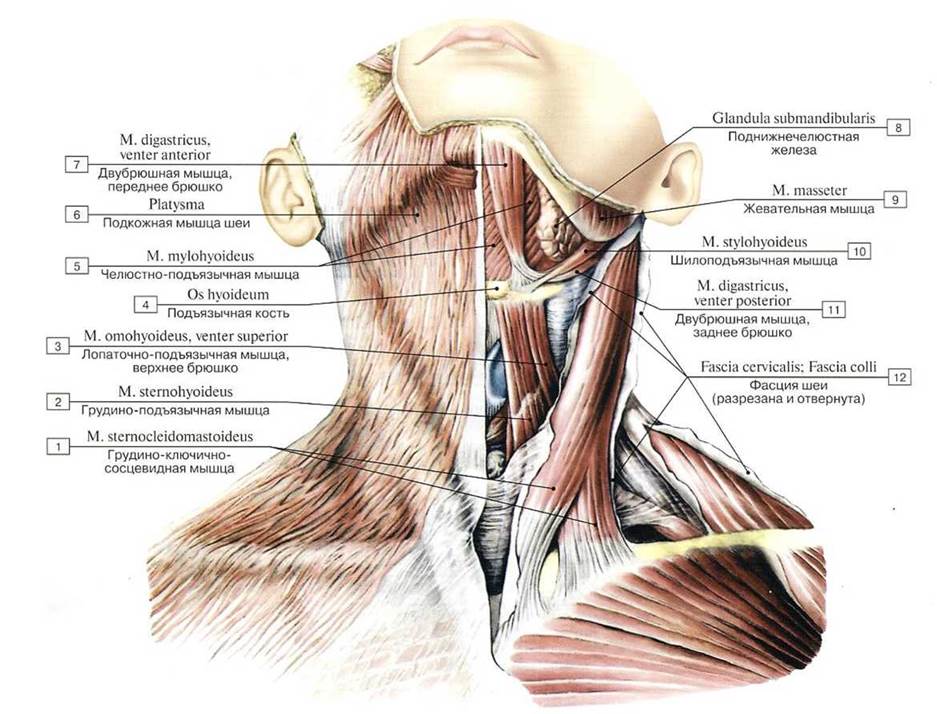 Подкожная мышца шеи и другие мышцы шеи, вид спереди;
1 — Sternocleidomastoid; 2 — Sternohyoid: 3 — Omohyoid, superior belly; 4 — Flyoid bone; 5 — Mylohyoid; 6 — Platysma; 7 — Digastric, anterior belly; 8 — Submandibular gland; 9 — Masseter; 10— Stylohyoid; 11 — Digastric, posterior belly; 12— Cervical fascia
Подкожная мышца шеи и другие мышцы шеи, вид спереди;
1 — Sternocleidomastoid; 2 — Sternohyoid: 3 — Omohyoid, superior belly; 4 — Flyoid bone; 5 — Mylohyoid; 6 — Platysma; 7 — Digastric, anterior belly; 8 — Submandibular gland; 9 — Masseter; 10— Stylohyoid; 11 — Digastric, posterior belly; 12— Cervical fascia
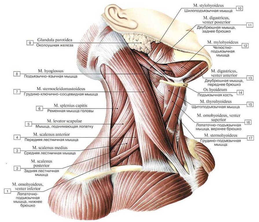 Грудино-ключично-сосцевидная мышца, надподъязычные и подподъязычные мышцы шеи, вид справа:
1 — Omohyoid, inferior belly; 2 — Scalenus posterior; Posterior scalene; 3 — Scalenus medius; Middle scalene: 4 — Scalenus anterior; Anterior scalene; 5 — Levator scapulae; 6 — Splenius capitis; 7 — Sternocleidomastoid; 8 — Hyoglossus; 9 — Parotid gland; 10 — Stylohyoid; 11 — Digastric, posterior belly; 12 — Mylohyoid; 13— Digastric, anterior belly; 14 — Hyoid bone; 15 —Thyrohyoid; 16 — Omohyoid, superior
belly; 17— Sternohyoid
Грудино-ключично-сосцевидная мышца, надподъязычные и подподъязычные мышцы шеи, вид справа:
1 — Omohyoid, inferior belly; 2 — Scalenus posterior; Posterior scalene; 3 — Scalenus medius; Middle scalene: 4 — Scalenus anterior; Anterior scalene; 5 — Levator scapulae; 6 — Splenius capitis; 7 — Sternocleidomastoid; 8 — Hyoglossus; 9 — Parotid gland; 10 — Stylohyoid; 11 — Digastric, posterior belly; 12 — Mylohyoid; 13— Digastric, anterior belly; 14 — Hyoid bone; 15 —Thyrohyoid; 16 — Omohyoid, superior
belly; 17— Sternohyoid
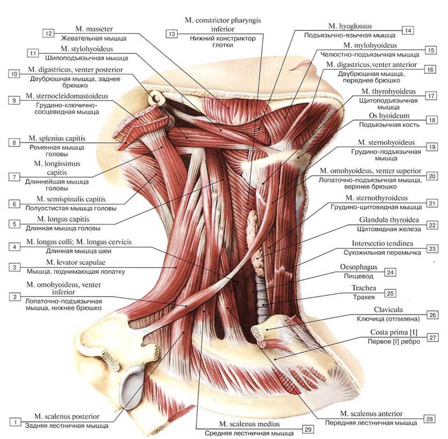 Мышцы шеи (мышцы, прикрепляющиеся к подъязычной кости), вид справа:
1 — Scalenus posterior; Posterior scalene; 2 — Omohyoid, inferior belly; 3 — Levator scapulae; 4 — Longus colli; 5 — Longus capitis; 6— Semispinalis capitis; 7— Longissimus capitis; 8 — Splenius capitis; 9 — Stemocleidomastoid; 10 — Digastric. posterior belly; 11 —Stylohyoid; 12 — Masseter; 13 — Inferior constrictor; 14 — Hyoglossus; 15 — Mylohyoid; 16 — Digastric, anterior belly; 17 — Thyrohyoid; 18 — Hyoid bone; 19 — Sternohyoid; 20 — Omohyoid, superior belly; 21 — Sternothyroid; 22 — Thyroid gland; 23 — Tendinous intersection; 24 —Oesophagus; 25 —Trachea; 26 — Clavicle: 27— First rib [1]; 28 — Scalenus anterior; Anterior scalene; 29— Scalenus medius; Middle scalene
Мышцы шеи (мышцы, прикрепляющиеся к подъязычной кости), вид справа:
1 — Scalenus posterior; Posterior scalene; 2 — Omohyoid, inferior belly; 3 — Levator scapulae; 4 — Longus colli; 5 — Longus capitis; 6— Semispinalis capitis; 7— Longissimus capitis; 8 — Splenius capitis; 9 — Stemocleidomastoid; 10 — Digastric. posterior belly; 11 —Stylohyoid; 12 — Masseter; 13 — Inferior constrictor; 14 — Hyoglossus; 15 — Mylohyoid; 16 — Digastric, anterior belly; 17 — Thyrohyoid; 18 — Hyoid bone; 19 — Sternohyoid; 20 — Omohyoid, superior belly; 21 — Sternothyroid; 22 — Thyroid gland; 23 — Tendinous intersection; 24 —Oesophagus; 25 —Trachea; 26 — Clavicle: 27— First rib [1]; 28 — Scalenus anterior; Anterior scalene; 29— Scalenus medius; Middle scalene
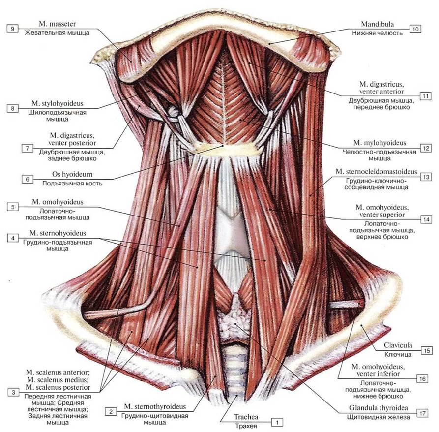 Поверхностные мышцы шеи, вид спереди. Подкожные и грудино-ключично-сосцевидные мышцы удалены:
1 — Trachea; 2 — Sternothyroid; 3 — Scalenus anterior; Anterior scalene; Scalenus medius; Middle scalene; Scalenus posterior; Posterior scalene; 4— Sternohyoid; 5— Omohyoid; 6 —Hyoid bone; 7— Digastric, posterior belly; 8 — Stylohyoid; 9 — Masseter; 10— Mandible; II- Digastric, anterior belly; 12 — Mylohyoid; 13 — Sternocleidomastoid; 14 — Omohyoid, superior belly; 15 — Clavicle; 16 — Omohyoid, inferior belly; 17— Thyroid gland
Поверхностные мышцы шеи, вид спереди. Подкожные и грудино-ключично-сосцевидные мышцы удалены:
1 — Trachea; 2 — Sternothyroid; 3 — Scalenus anterior; Anterior scalene; Scalenus medius; Middle scalene; Scalenus posterior; Posterior scalene; 4— Sternohyoid; 5— Omohyoid; 6 —Hyoid bone; 7— Digastric, posterior belly; 8 — Stylohyoid; 9 — Masseter; 10— Mandible; II- Digastric, anterior belly; 12 — Mylohyoid; 13 — Sternocleidomastoid; 14 — Omohyoid, superior belly; 15 — Clavicle; 16 — Omohyoid, inferior belly; 17— Thyroid gland
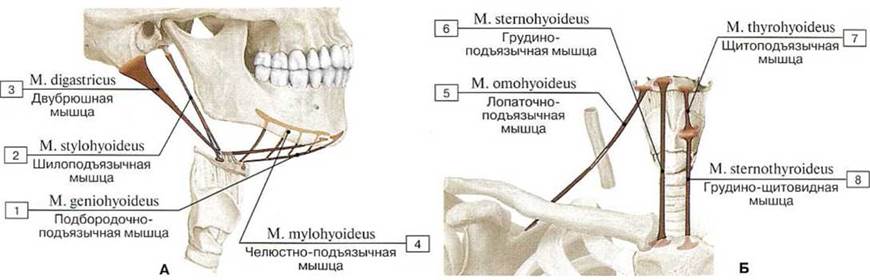 Места начала и прикрепления (А — надподъязычных мышц, Б — подподъязычных мышц) (схемы):
1 — Geniohyoid; 2— Stylohyoid; 3 — Digastric; 4— Mylohyoid; 5— Omohyoid; 6 — Sternohyoid; 7 — Thyrohyoid; 8 — Sternothyroid
Места начала и прикрепления (А — надподъязычных мышц, Б — подподъязычных мышц) (схемы):
1 — Geniohyoid; 2— Stylohyoid; 3 — Digastric; 4— Mylohyoid; 5— Omohyoid; 6 — Sternohyoid; 7 — Thyrohyoid; 8 — Sternothyroid
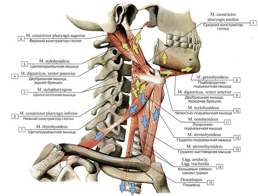 Направления тяги мышц, прикрепляющихся к подъязычной кости (схема)
1 — Thyrohyoid; 2 — Inferior constrictor; 3 — Stylopharyngeus; 4 — Digastric, posterior belly; 5 — Stylohyoid; 6 — Superior constrictor; 7— Middle constrictor; 8 — Geniohyoid; 9 — Digastric, anterior belly; 10 — Mylohyoid; 11 — Omohyoid; 12 — Sternohyoid;13 — Sternothyroid; 14 — Anular ligaments; 15 — Oesophagus
Направления тяги мышц, прикрепляющихся к подъязычной кости (схема)
1 — Thyrohyoid; 2 — Inferior constrictor; 3 — Stylopharyngeus; 4 — Digastric, posterior belly; 5 — Stylohyoid; 6 — Superior constrictor; 7— Middle constrictor; 8 — Geniohyoid; 9 — Digastric, anterior belly; 10 — Mylohyoid; 11 — Omohyoid; 12 — Sternohyoid;13 — Sternothyroid; 14 — Anular ligaments; 15 — Oesophagus
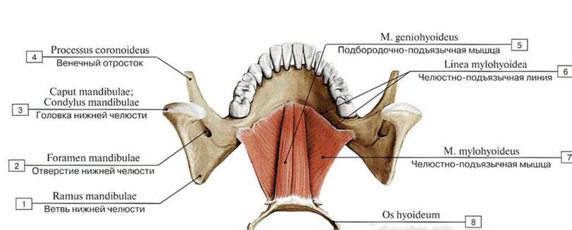 Челюстно-подъязычные и подбородочно-подъязычные мышцы, вид сверху:
1 — Ramus of mandible; 2 — Mandibular foramen; 3 — Head of mandible; 4 — Coronoid process; 5 — Geniohyoid; 6 — Mylohyoid line;
7— Mylohyoid; 8— Hyoid bone
Челюстно-подъязычные и подбородочно-подъязычные мышцы, вид сверху:
1 — Ramus of mandible; 2 — Mandibular foramen; 3 — Head of mandible; 4 — Coronoid process; 5 — Geniohyoid; 6 — Mylohyoid line;
7— Mylohyoid; 8— Hyoid bone
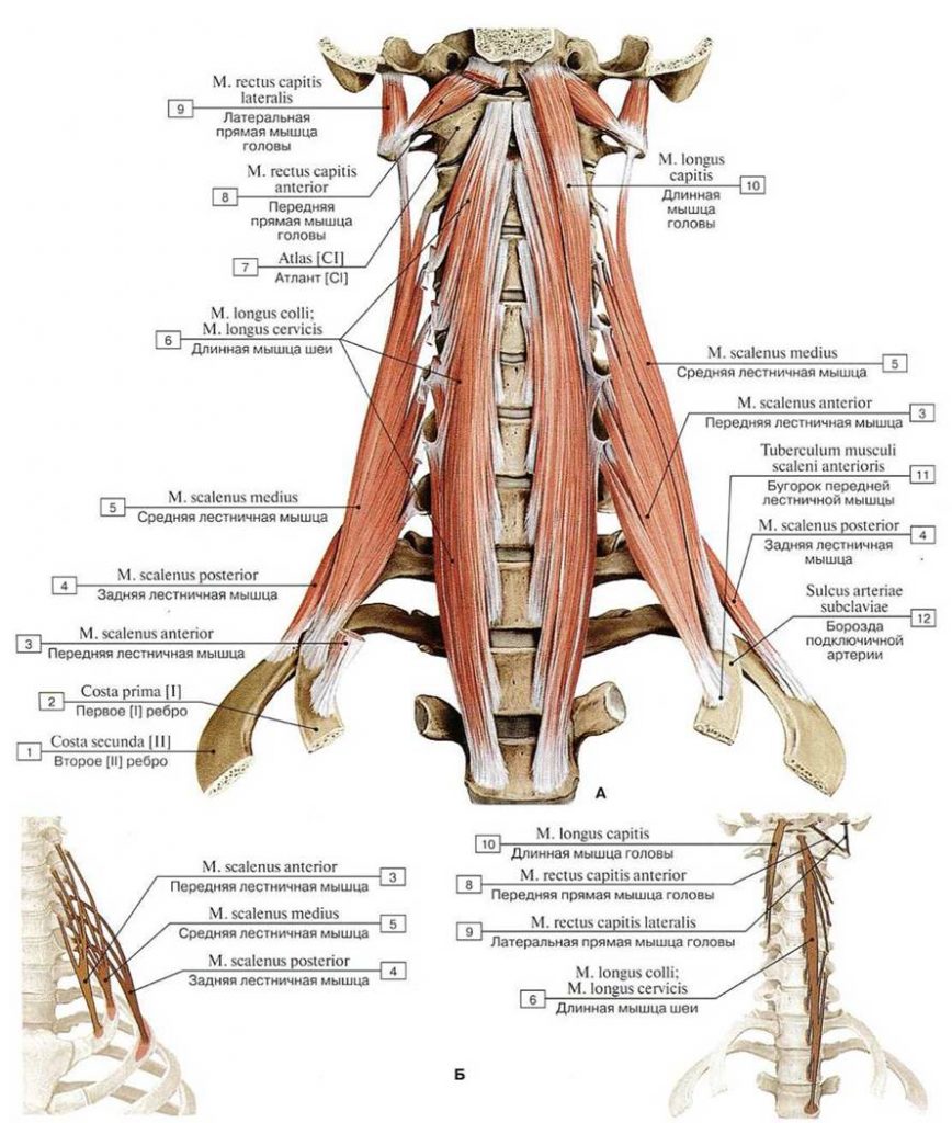 Глубокие мышцы шеи, вид спереди (А — общий вид, Б — места начала и прикрепления глубоких мышц шеи (схемы)):
1 — Second rib [II]; 2 — First rib [I]; 3 — Scalenus anterior; Anterior scalene; 4 —-Scalenus posterior; Posterior scalene; 5— Scalenus medius; Middle scalene; 6 — Longus colli; 7 — Atlas [CI]; 8— Rectus capitis anterior; 9 — Rectus capitis lateralis; 10 — Longus capitis; 11 — Scalene tubercle; 12— Groove for subclavian artery
Глубокие мышцы шеи, вид спереди (А — общий вид, Б — места начала и прикрепления глубоких мышц шеи (схемы)):
1 — Second rib [II]; 2 — First rib [I]; 3 — Scalenus anterior; Anterior scalene; 4 —-Scalenus posterior; Posterior scalene; 5— Scalenus medius; Middle scalene; 6 — Longus colli; 7 — Atlas [CI]; 8— Rectus capitis anterior; 9 — Rectus capitis lateralis; 10 — Longus capitis; 11 — Scalene tubercle; 12— Groove for subclavian artery
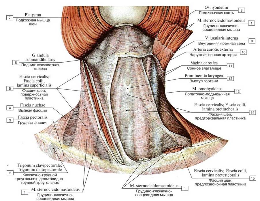
 Пластинки шейной фасции, вид спереди:
1 — Stemohyoid; 2 — Invcsting layer; 3 — Parotid gland; 4 — Mandible; 5 — Sternocleidomastoid; 6 — Carotid sheath; 7— Trapezius;
8 — Prevertebral laver; 9 — Clavicle
Пластинки шейной фасции, вид спереди:
1 — Stemohyoid; 2 — Invcsting layer; 3 — Parotid gland; 4 — Mandible; 5 — Sternocleidomastoid; 6 — Carotid sheath; 7— Trapezius;
8 — Prevertebral laver; 9 — Clavicle
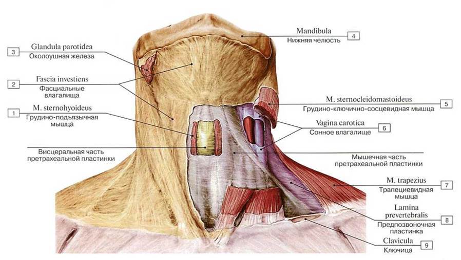
 Фасции шеи, вид спереди:
1 — Sternocleidomastoid; 2 — Clavipectoral triangle; Dcltopectoral triangle; 3 — Pectoral fascia; 4 — Nuchal fascia; 5 — Cervical fascia, inves- ting laver; superficial layer; 6 — Submandibulargland; 7— Platysma; 8 — Hyoid bone; 9 —Internal jugularvein; 10 — External carotid artery; 11 — Carotid sheath; 12 — Laryngeal prominence; 13 — Omohyoid; 14 — Cervical fascia, pretracheal layer; 15 — Cervical fascia, prevertebral layer
Фасции шеи, вид спереди:
1 — Sternocleidomastoid; 2 — Clavipectoral triangle; Dcltopectoral triangle; 3 — Pectoral fascia; 4 — Nuchal fascia; 5 — Cervical fascia, inves- ting laver; superficial layer; 6 — Submandibulargland; 7— Platysma; 8 — Hyoid bone; 9 —Internal jugularvein; 10 — External carotid artery; 11 — Carotid sheath; 12 — Laryngeal prominence; 13 — Omohyoid; 14 — Cervical fascia, pretracheal layer; 15 — Cervical fascia, prevertebral layer
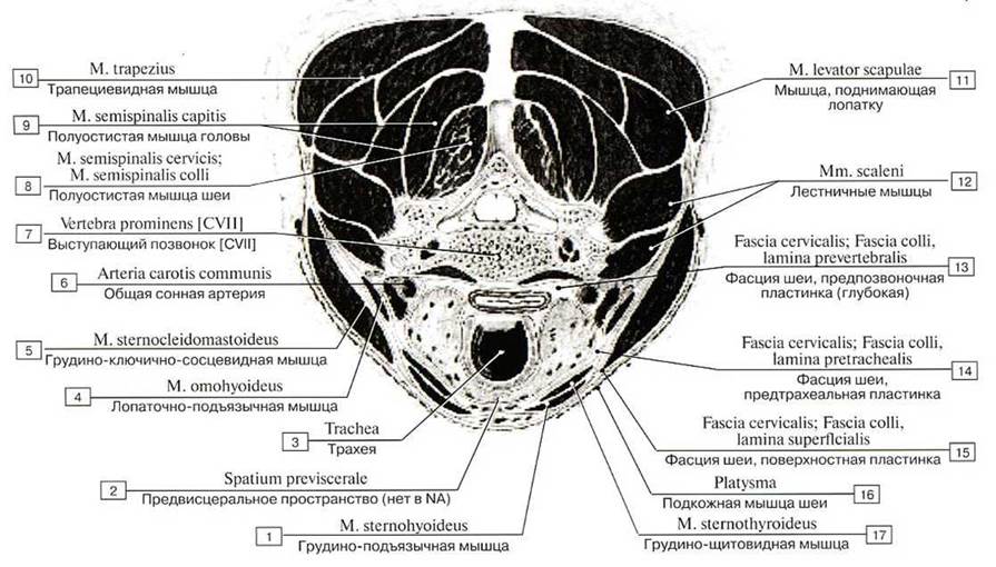 Мышцы и фасции шеи на поперечном разрезе. Разрез сделан на уровне VII шейного позвонка и шитовидной
железы:
1 — Slernohyoid; 2 — Previsceral space; 3 — Trachea; 4 — Omohyoid; 5 — Sternocleidomastoid; 6 — Common carotid artery; 7— Vertebra prominens [CVII]; 8 — Semispinalis cervicis; 9 —Semispinalis capitis; 10 — Trapezius; 11 — Levator scapulae; 12 — Scalenus; 13 — Cervical fascia, prevertebral laver; 14 — Cervical fascia, pretracheal laver; 15 — Cervical fascia, investing layer; superficial layer; 16 — Platysma; 17— Sternothyroid
Мышцы и фасции шеи на поперечном разрезе. Разрез сделан на уровне VII шейного позвонка и шитовидной
железы:
1 — Slernohyoid; 2 — Previsceral space; 3 — Trachea; 4 — Omohyoid; 5 — Sternocleidomastoid; 6 — Common carotid artery; 7— Vertebra prominens [CVII]; 8 — Semispinalis cervicis; 9 —Semispinalis capitis; 10 — Trapezius; 11 — Levator scapulae; 12 — Scalenus; 13 — Cervical fascia, prevertebral laver; 14 — Cervical fascia, pretracheal laver; 15 — Cervical fascia, investing layer; superficial layer; 16 — Platysma; 17— Sternothyroid
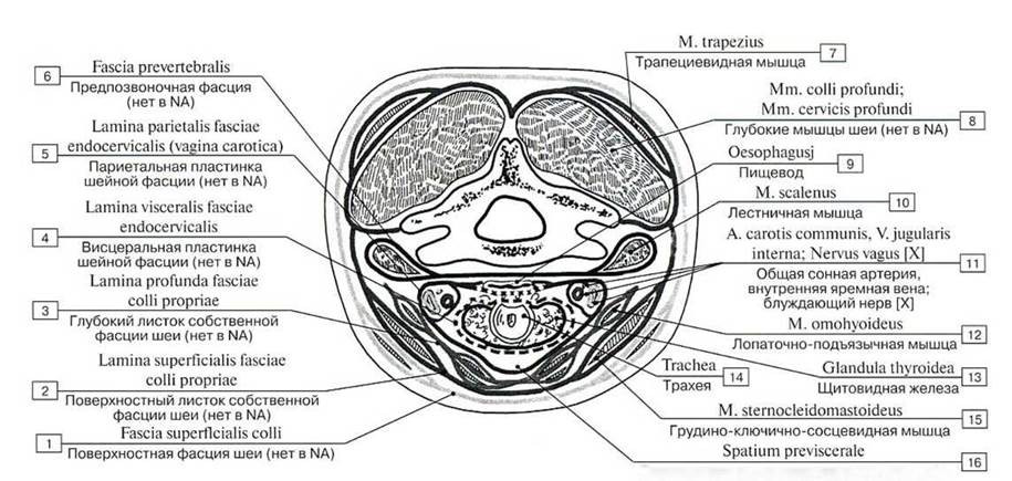 Фасции шеи, горизонтальный разрез (схема); 1 — Superficia! fascia of neck; 2 — Superficial layer of cervical fascia; 3 — Deep layer of cervical fascia; 4 — Visceral layer of endocervi- cal fascia; 5 — Parietal layer of endocervical fascia; 6 — Prevertebral fascia; 7 — Trapezius; 8 — Deep muscles of neck; 9 — Oesophagus; 10 — Scalenus; 11 — Common carotid artery, Internal jugular vein: Vagus nerve [X]; 12 — Omohyoid; 13 — Thyroid gland; 14 — Trachea; 15 — Stemoeleidomastoid; 16 — Prcvisceral space (по Шевкуненко)
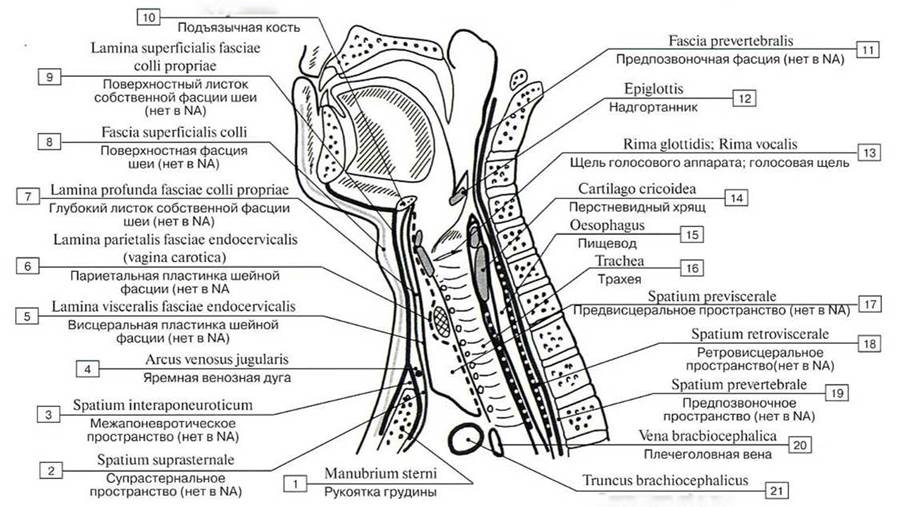 Фасции и клетчаточные пространства шеи, сагиттальный разрез (схема):
1 — Manubrium of sternum; 2 — Suprasternal space; 3 — Interaponeurotic space; 4 — Jugular venous arch; 5 — Visceral layer of endocervical fascia; 6 — Parietal layer of endocervical fascia; 7 — Deep layer of cervical fascia; 8 — Superficial fascia of neck; 9 — Superficial layer of cervical fascia: 10 — Hyoid bone; 11— Prevertebral fascia; 12— Epiglottis; 13 — Rima glottidis; 14 — Cricoid cartilage; 15— Oesophagus; 16 — Trachea; 17 — Previsceral space; 18 — Retrovisceral space; 19— Prevertebral space; 20 — Brachiocephalic vein; 21 — Brachiocephalic trunk (по Шевкуненко)
Фасции и клетчаточные пространства шеи, сагиттальный разрез (схема):
1 — Manubrium of sternum; 2 — Suprasternal space; 3 — Interaponeurotic space; 4 — Jugular venous arch; 5 — Visceral layer of endocervical fascia; 6 — Parietal layer of endocervical fascia; 7 — Deep layer of cervical fascia; 8 — Superficial fascia of neck; 9 — Superficial layer of cervical fascia: 10 — Hyoid bone; 11— Prevertebral fascia; 12— Epiglottis; 13 — Rima glottidis; 14 — Cricoid cartilage; 15— Oesophagus; 16 — Trachea; 17 — Previsceral space; 18 — Retrovisceral space; 19— Prevertebral space; 20 — Brachiocephalic vein; 21 — Brachiocephalic trunk (по Шевкуненко)
Страницы:
1 2 3 4 5 6 7 8 9 10 11 12 13 14 15 16 17 18