Перейти к содержимому
опография фасций и клетчаточных пространств нижней конечности
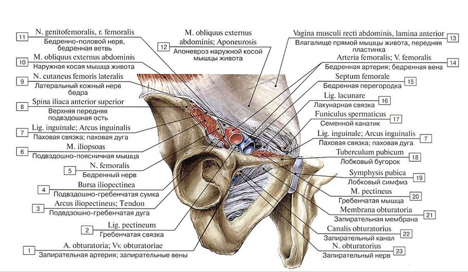 Мышечная и сосудистая лакуны:
1 — Obturator artery; Obturator veins; 2 — Pectineal ligament; 3 — Iliopectineal arch; Tendon; 4 — Iliopectincal bursa; 5 — Femoral петле; 6 —- Iliopsoas; 7 — Inguinal ligament; 8 — Anterior superior iliac spine; 9 — Lateral cutaneous nerve of thigh; Lateral femoral cutaneous nerve; 10 — External oblique; 11 — Genitofemoral nerve, femoral branch; 12 — External oblique; Aponeurosis; 13 — Rectus sheath, anterior layer; 14 — Femoral artery; Femoral vein; 15 — Femoral septum; 16 — Lacunar ligament; 17 — Spermatic cord; 18 — Pubic tubercle; 19 — Pubic symphysis; 20 — Pectineus; 21 — Obturator membrane; 22 — Obturator canal; 23 — Obturator nerve
Мышечная и сосудистая лакуны:
1 — Obturator artery; Obturator veins; 2 — Pectineal ligament; 3 — Iliopectineal arch; Tendon; 4 — Iliopectincal bursa; 5 — Femoral петле; 6 —- Iliopsoas; 7 — Inguinal ligament; 8 — Anterior superior iliac spine; 9 — Lateral cutaneous nerve of thigh; Lateral femoral cutaneous nerve; 10 — External oblique; 11 — Genitofemoral nerve, femoral branch; 12 — External oblique; Aponeurosis; 13 — Rectus sheath, anterior layer; 14 — Femoral artery; Femoral vein; 15 — Femoral septum; 16 — Lacunar ligament; 17 — Spermatic cord; 18 — Pubic tubercle; 19 — Pubic symphysis; 20 — Pectineus; 21 — Obturator membrane; 22 — Obturator canal; 23 — Obturator nerve
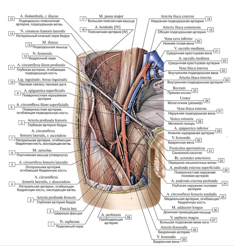 Переход наружной подвздошной артерии в бедренную артерию:
1 — Saphenous nerve; 2 — Fascia lata; 3 — Deep artery of thigh; 4 — Lateral circumflex femoral artery, descending branch; 5 — Lateral circumflex femoral artery: 6 — Sartorius; 7— Lateral circumflex femoral artery, ascending branch; 8 — Superficial circumflex iliac artery; 9 — Superficial epigastric artery; 10 — Inguinal ligament; 11 — Deep circumflex iliac artery; 12 — Femoral nerve; 13 — Iliacus; 14 — Lateral cutaneous nerve of thigh; Lateral femoral cutaneous nerve; 15 — Iliolumbar artery, iliacus branch; 16 — Lumbar artery [IV]; 17— Psoas major; 18 — External iliac artery; 19 — Common iliac artery; 20 — Inferior vena cava; 21 — Median sacral vein; 22 — Median sacral artery; 23 — Internal iliac vein; 24 — Internal iliac artery; 25 — Rectum; 26 — Ureter; 27 — External iliac vein; 28 — Urinary bladder; 29 — Inferior epigastric artery; 30 — Femoral vein; 31 — Spermatic cord; 32 — Anterior scrotal branches; 33 — Superficial external pudendal artery; 34 — Deep external pudendal artery; 35 — Medial circumflex femoral artery; 36 — Adductor longus; 37— Great saphenous vein; Long saphenous vein; 38 — Femoral artery; 39 — Perforating artery
Переход наружной подвздошной артерии в бедренную артерию:
1 — Saphenous nerve; 2 — Fascia lata; 3 — Deep artery of thigh; 4 — Lateral circumflex femoral artery, descending branch; 5 — Lateral circumflex femoral artery: 6 — Sartorius; 7— Lateral circumflex femoral artery, ascending branch; 8 — Superficial circumflex iliac artery; 9 — Superficial epigastric artery; 10 — Inguinal ligament; 11 — Deep circumflex iliac artery; 12 — Femoral nerve; 13 — Iliacus; 14 — Lateral cutaneous nerve of thigh; Lateral femoral cutaneous nerve; 15 — Iliolumbar artery, iliacus branch; 16 — Lumbar artery [IV]; 17— Psoas major; 18 — External iliac artery; 19 — Common iliac artery; 20 — Inferior vena cava; 21 — Median sacral vein; 22 — Median sacral artery; 23 — Internal iliac vein; 24 — Internal iliac artery; 25 — Rectum; 26 — Ureter; 27 — External iliac vein; 28 — Urinary bladder; 29 — Inferior epigastric artery; 30 — Femoral vein; 31 — Spermatic cord; 32 — Anterior scrotal branches; 33 — Superficial external pudendal artery; 34 — Deep external pudendal artery; 35 — Medial circumflex femoral artery; 36 — Adductor longus; 37— Great saphenous vein; Long saphenous vein; 38 — Femoral artery; 39 — Perforating artery
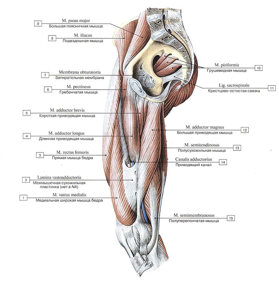 Приводящий канал и приводящая мышца бедра, правого, вид с медиальной стороны:
1 — Vastus medialis; 2 — Vastoadductore plate; 3 — Rectus femoris; 4 — Adductor longus; 5 — Adductor brevis; 6 — Pectineus; 7 — Obturator membrane; 8 — Iliacus; 9 — Psoas major; 10 — Piriformis; 11 — Sacrospinous ligament; 12 — Adductor magnus; 13 — Semitendinosus; 14 — Adductor canal; 15 — Semimembranosus
Приводящий канал и приводящая мышца бедра, правого, вид с медиальной стороны:
1 — Vastus medialis; 2 — Vastoadductore plate; 3 — Rectus femoris; 4 — Adductor longus; 5 — Adductor brevis; 6 — Pectineus; 7 — Obturator membrane; 8 — Iliacus; 9 — Psoas major; 10 — Piriformis; 11 — Sacrospinous ligament; 12 — Adductor magnus; 13 — Semitendinosus; 14 — Adductor canal; 15 — Semimembranosus
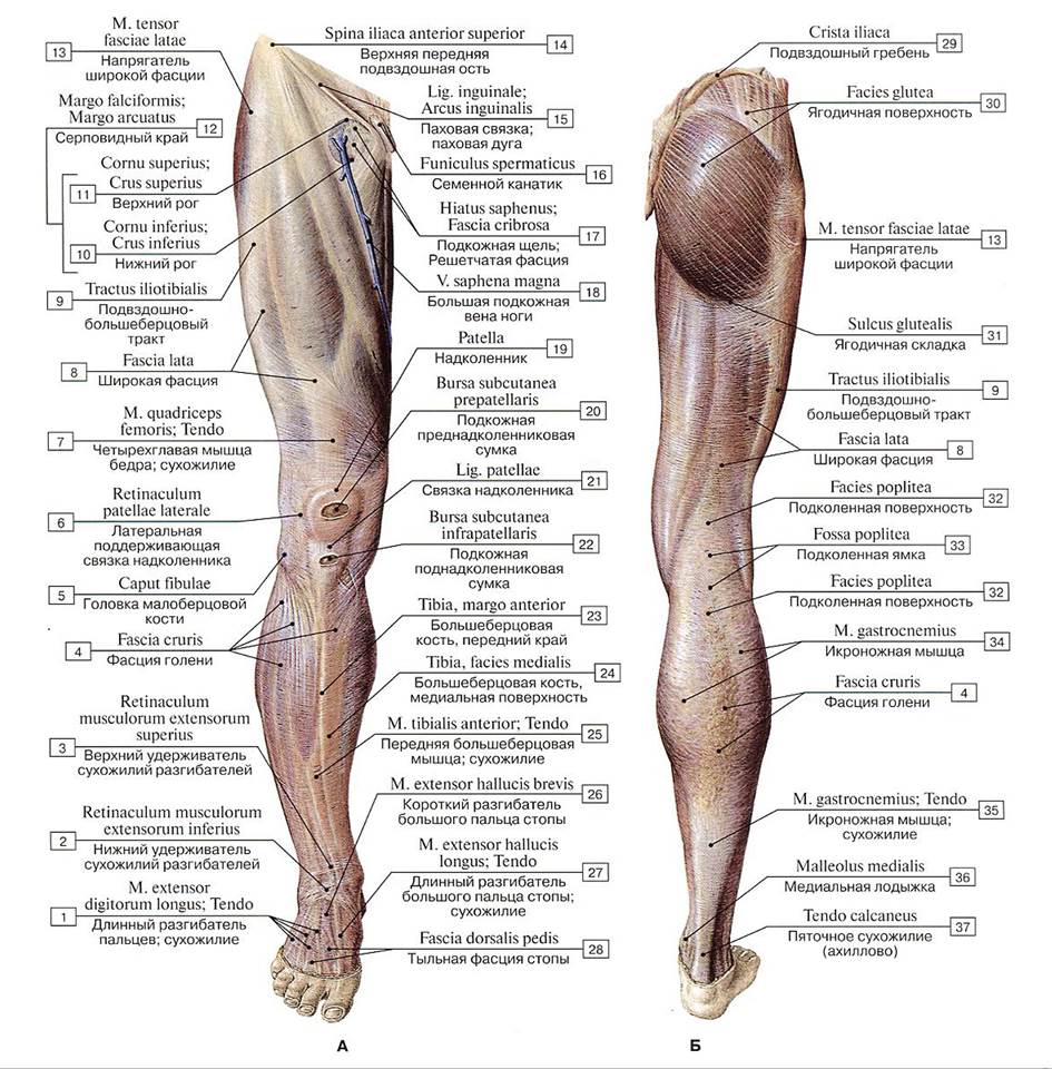 Фасции нижней конечности, правой (А — вид спереди, Б — вид сзади):
1 — Extensor digitorum longus; Tendon; 2 — Inferior extensor retinaculum; 3 — Superior extensor retinaculum; 4 — Deep fascia of leg; 5- Head of fibula; 6— Lateral patellar retinaculum; 7— Quadriceps femoris; Tendon; 8 — Fascia lata; 9— Iliotibial tract; 10 — Inferior horn; 11 — Superior horn; 12=10+11 — Falciform margin; 13 — Tensor fasciae latae; Tensor of fascia lata; 14 — Anterior superior iliac spine; 15— Inguinal ligament; 16— Spermatic cord; 17—Saphenous opening; Cribriform fascia; 18 — Great saphenous vein; Long saphenous vein; 19 — Patella; 20 — Subcutaneous prepatellar bursa; 21 — Patellar ligament; 22 — Subcutaneous infrapatellar bursa; 23 — Tibia, anterior border; 24 — Tibia, medial surface; 25 — Tibialis anterior; Tendon; 26 —- Extensor hallueis brevis; 27— Extensor hallueis longus; Tendon; 28 —- Dorsal fascia of foot; 29 — Iliac crest; 30 — Gluteal surface; 31 — Gluteal fold; 32 — Popliteal surface; 33 — Popliteal fossa; 34 — Gastrocnemius;
35 — Gastrocnemius; Tendon; 36— Medial malleolus; 37— Calcaneal tendon
Фасции нижней конечности, правой (А — вид спереди, Б — вид сзади):
1 — Extensor digitorum longus; Tendon; 2 — Inferior extensor retinaculum; 3 — Superior extensor retinaculum; 4 — Deep fascia of leg; 5- Head of fibula; 6— Lateral patellar retinaculum; 7— Quadriceps femoris; Tendon; 8 — Fascia lata; 9— Iliotibial tract; 10 — Inferior horn; 11 — Superior horn; 12=10+11 — Falciform margin; 13 — Tensor fasciae latae; Tensor of fascia lata; 14 — Anterior superior iliac spine; 15— Inguinal ligament; 16— Spermatic cord; 17—Saphenous opening; Cribriform fascia; 18 — Great saphenous vein; Long saphenous vein; 19 — Patella; 20 — Subcutaneous prepatellar bursa; 21 — Patellar ligament; 22 — Subcutaneous infrapatellar bursa; 23 — Tibia, anterior border; 24 — Tibia, medial surface; 25 — Tibialis anterior; Tendon; 26 —- Extensor hallueis brevis; 27— Extensor hallueis longus; Tendon; 28 —- Dorsal fascia of foot; 29 — Iliac crest; 30 — Gluteal surface; 31 — Gluteal fold; 32 — Popliteal surface; 33 — Popliteal fossa; 34 — Gastrocnemius;
35 — Gastrocnemius; Tendon; 36— Medial malleolus; 37— Calcaneal tendon
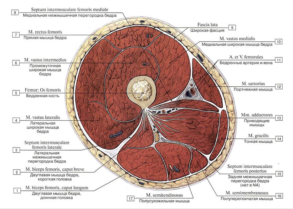 Поперечный распил бедра на уровне средней трети:
1— Biceps femoris, long head; 2 —- Biceps femoris, short head; 3 — Lateral femoral intermuscular septum; 4 — Vastus lateralis; 5 — Femur; Thigh bone: 6 —- Vastus intermedius; 7— Rectus femoris; 8 — Medial femoral intermuscular septum; 9 — Fascia lata; 10— Vastus medialis; 11 — Femoral artery and vein; 12—Sartorius; 13 — Adductores; 14— Gracilis; 15—Posterior femoral intermuscular septum; 16— Semimem branosus; 17— Semitendinosus
Поперечный распил бедра на уровне средней трети:
1— Biceps femoris, long head; 2 —- Biceps femoris, short head; 3 — Lateral femoral intermuscular septum; 4 — Vastus lateralis; 5 — Femur; Thigh bone: 6 —- Vastus intermedius; 7— Rectus femoris; 8 — Medial femoral intermuscular septum; 9 — Fascia lata; 10— Vastus medialis; 11 — Femoral artery and vein; 12—Sartorius; 13 — Adductores; 14— Gracilis; 15—Posterior femoral intermuscular septum; 16— Semimem branosus; 17— Semitendinosus
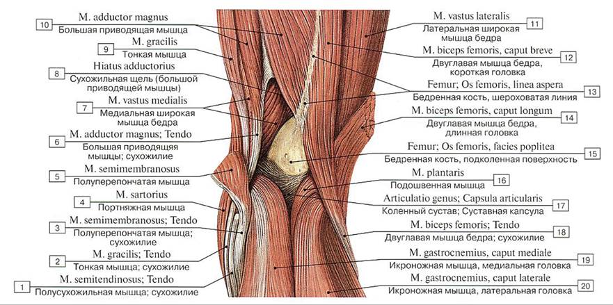 Подколенная ямка правой нижней конечности, вид сзади:
1 — Semitendinosus; Tendon; 2 — Gracilis: Tendon; 3 — Semimembranosus; Tendon; 4 — Sartorius; 5 — Semimembranosus; 6 — Adductor magnus; Tendon; 7 — Vastus medialis; 8— Adductor hiatus; 9— Gracilis; 10— Adductor magnus; 11 — Vastus lateralis; 12— Biceps femoris, short head; 13— Femur; Thigh bone, lineaaspera; 14— Biceps femoris, longhead: 15— Femur; Thigh bone, popliteal surface; 16 — Plantaris; 17 — Knee joint; Joint capsule; Articular capsule; 18 — Biceps femoris; Tendon; 19 — Gastrocnemius, medial head; 20— Gastrocnemius, lateral head
Подколенная ямка правой нижней конечности, вид сзади:
1 — Semitendinosus; Tendon; 2 — Gracilis: Tendon; 3 — Semimembranosus; Tendon; 4 — Sartorius; 5 — Semimembranosus; 6 — Adductor magnus; Tendon; 7 — Vastus medialis; 8— Adductor hiatus; 9— Gracilis; 10— Adductor magnus; 11 — Vastus lateralis; 12— Biceps femoris, short head; 13— Femur; Thigh bone, lineaaspera; 14— Biceps femoris, longhead: 15— Femur; Thigh bone, popliteal surface; 16 — Plantaris; 17 — Knee joint; Joint capsule; Articular capsule; 18 — Biceps femoris; Tendon; 19 — Gastrocnemius, medial head; 20— Gastrocnemius, lateral head
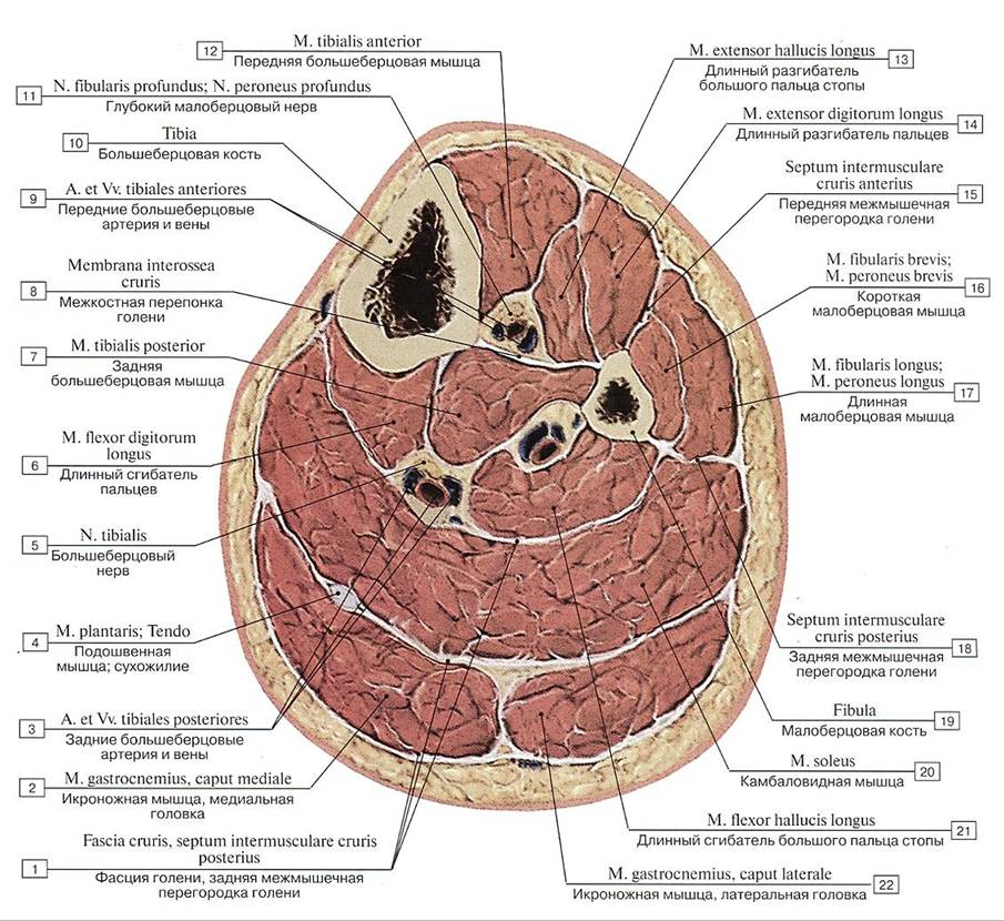 Поперечный распил голени на уровне верхней трети:
1 — Deep fascia of leg, posterior intermuscular septum of leg; 2 — Gastrocnemius, medial head; 3 — Posterior tibial artery and veins; 4 — Plantaris; Tendon; 5 — Tibial nerve; 6 — Flexor digitorum longus; 7— Tibialis posterior; 8 — Interosseous membrane ofleg; 9 — Anterior tibial artery and veins; 10 —- Tibia; 11 — Deep fibular nerve; Deep peroneal nerve; 12 — Tibialis anterior; 13 — Extensor hallucis longus; 14 — Extensor digitorum longus; 15 — Anterior intermuscular septum of leg; 16 — Fibularis brevis; Peroneus brevis; 17 — Fibularis longus; Peroneus longus; 18 — Posterior intermuscular septum of leg; 19 — Fibula; 20 — Soleus; 21 — Flexor hallucis longus; 22 — Gastrocnemius, lateral head
Поперечный распил голени на уровне верхней трети:
1 — Deep fascia of leg, posterior intermuscular septum of leg; 2 — Gastrocnemius, medial head; 3 — Posterior tibial artery and veins; 4 — Plantaris; Tendon; 5 — Tibial nerve; 6 — Flexor digitorum longus; 7— Tibialis posterior; 8 — Interosseous membrane ofleg; 9 — Anterior tibial artery and veins; 10 —- Tibia; 11 — Deep fibular nerve; Deep peroneal nerve; 12 — Tibialis anterior; 13 — Extensor hallucis longus; 14 — Extensor digitorum longus; 15 — Anterior intermuscular septum of leg; 16 — Fibularis brevis; Peroneus brevis; 17 — Fibularis longus; Peroneus longus; 18 — Posterior intermuscular septum of leg; 19 — Fibula; 20 — Soleus; 21 — Flexor hallucis longus; 22 — Gastrocnemius, lateral head
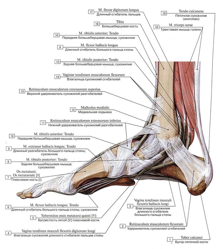 Удерживатели сухожилий стопы, правой, вид с медиальной стороны:
1 — Calcaneal tuber; 2 — Flexor retinaculum; 3 —Tendinous sheath oflong flexor muscle ofgrcat toe; 4 —Tendinous sheath oflong flexor muscle of toes; 5 — Tuberosity of fifth metatarsal bone [V]; 6 — Flexor hallucis longus; Tendon; 7— Metatarsal [1]; 8 — Tibialis posterior; Tendon; 9 — Extensor hallucis longus: Tendon; 10 — Tibialis anterior; Tendon; 11 — Inferior extensor retinaculum; 12 — Medial malleolus; 13 — Superior extensor retinaculum; 14 — Tendinous sheaths of flexor muscles; 15 — Tibialis posterior; Tendon; 16 — Tibia; 17— Flexor digitorum longus; 18 — Triceps surae; 19 — Calcaneal tendon
Удерживатели сухожилий стопы, правой, вид с медиальной стороны:
1 — Calcaneal tuber; 2 — Flexor retinaculum; 3 —Tendinous sheath oflong flexor muscle ofgrcat toe; 4 —Tendinous sheath oflong flexor muscle of toes; 5 — Tuberosity of fifth metatarsal bone [V]; 6 — Flexor hallucis longus; Tendon; 7— Metatarsal [1]; 8 — Tibialis posterior; Tendon; 9 — Extensor hallucis longus: Tendon; 10 — Tibialis anterior; Tendon; 11 — Inferior extensor retinaculum; 12 — Medial malleolus; 13 — Superior extensor retinaculum; 14 — Tendinous sheaths of flexor muscles; 15 — Tibialis posterior; Tendon; 16 — Tibia; 17— Flexor digitorum longus; 18 — Triceps surae; 19 — Calcaneal tendon
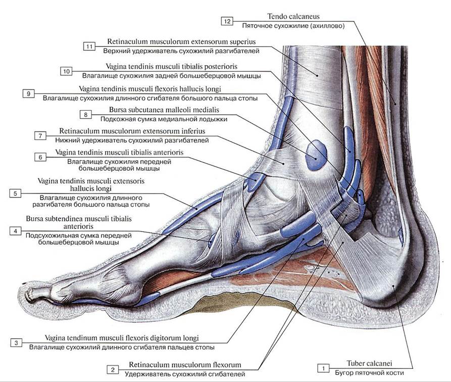 Синовиальные влагалища сухожилий стопы, правой, вид с медиальной стороны:
1 — Calcaneal tuber; 2 — Flexor retinaculum; 2 — Tendinous sheath of flexor digitorum longus; 4 — Subtendinous bursa of tibialis anterior; 5 — Tendinous sheath of extensor hallucis longus; 6 — Tendinous sheath of tibialis anterior; 7 — Inferior extensor retinaculum; 8 — Subcutaneous bursa of medial malleolus; 9 — Tendinous sheath of flexor hallucis longus; 10 — Tendinous sheath of tibialis posterior;
11 — Superior extensor retinaculum; 12 — Calcaneal tendon
Синовиальные влагалища сухожилий стопы, правой, вид с медиальной стороны:
1 — Calcaneal tuber; 2 — Flexor retinaculum; 2 — Tendinous sheath of flexor digitorum longus; 4 — Subtendinous bursa of tibialis anterior; 5 — Tendinous sheath of extensor hallucis longus; 6 — Tendinous sheath of tibialis anterior; 7 — Inferior extensor retinaculum; 8 — Subcutaneous bursa of medial malleolus; 9 — Tendinous sheath of flexor hallucis longus; 10 — Tendinous sheath of tibialis posterior;
11 — Superior extensor retinaculum; 12 — Calcaneal tendon
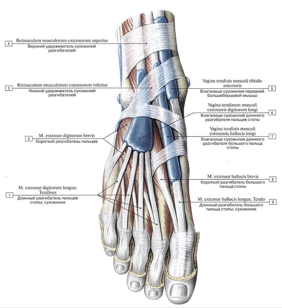 Синовиальные влагалища сухожилий мышц-разгибателей стопы, правой, и ее пальцев, вил спереди и сверху:
1 — Extensor digitorum longus; Tendons; 2 — Extensor digitorum brevis; 3 — Inferior extensor retinaculum; 4 — Superior extensor retinaculum; 5 — Tendinous sheath of tibialis anterior; 6 — Tendinous sheath of extensor digitorum longus; 7 — Tendinous sheath of extensor halluces longus; 8— Extensor hallucis brevis; 9 — Extensor hallucis longus; Tendon
Синовиальные влагалища сухожилий мышц-разгибателей стопы, правой, и ее пальцев, вил спереди и сверху:
1 — Extensor digitorum longus; Tendons; 2 — Extensor digitorum brevis; 3 — Inferior extensor retinaculum; 4 — Superior extensor retinaculum; 5 — Tendinous sheath of tibialis anterior; 6 — Tendinous sheath of extensor digitorum longus; 7 — Tendinous sheath of extensor halluces longus; 8— Extensor hallucis brevis; 9 — Extensor hallucis longus; Tendon
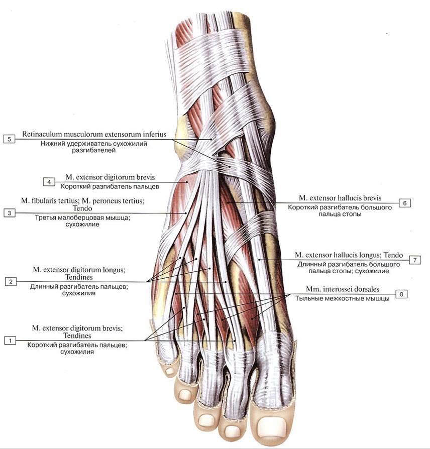 Сухожилия разгибателей и тыльные мышцы стопы, правой, вид сверху:
1 — Extensor digitorum brevis; Tendons; 2 — Extensor digitorum longus; Tendons; 3 — Fibularis tertius; Peroneus tertius: Tendon; 4 — Extensor digitorum brevis; 5 — Inferior extensor retinaculum; 6 — Extensor hallucis brevis; 7 — Extensor hallucis longus; Tendon; 8 — Dorsal interossei
Сухожилия разгибателей и тыльные мышцы стопы, правой, вид сверху:
1 — Extensor digitorum brevis; Tendons; 2 — Extensor digitorum longus; Tendons; 3 — Fibularis tertius; Peroneus tertius: Tendon; 4 — Extensor digitorum brevis; 5 — Inferior extensor retinaculum; 6 — Extensor hallucis brevis; 7 — Extensor hallucis longus; Tendon; 8 — Dorsal interossei
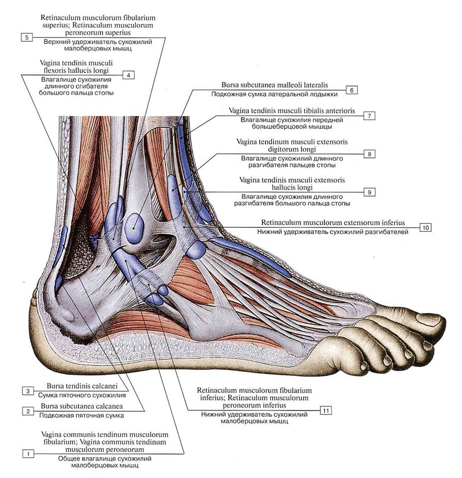 Синовиальные влагалища сухожилий стопы, правой, вид с латеральной стороны:
1 — Common tendinous sheath of fibulares; Common tendinous sheath of peronei; 2 — Subcutaneous calcaneal bursa; 3 —- Bursa of tendo calcaneus; Bursa of calcaneal tendon; Retrocalcancal bursa; 4 — Tendinous sheath of flexor hallucis longus; 5 — Superior fibular retinaculum; Superior peroneal retinaculum; 6 — Subcutaneous bursa of lateral malleolus; 7 — Tendinous sheath of tibialis anterior; 8 — Tendinous sheath of extensor digitorum longus; 9 — Tendinous sheath of extensor hallucis longus; 10 — Inferior extensor retinaculum; 11 — Inferior fibular retinaculum; Inferior peroneal retinaculum
Синовиальные влагалища сухожилий стопы, правой, вид с латеральной стороны:
1 — Common tendinous sheath of fibulares; Common tendinous sheath of peronei; 2 — Subcutaneous calcaneal bursa; 3 —- Bursa of tendo calcaneus; Bursa of calcaneal tendon; Retrocalcancal bursa; 4 — Tendinous sheath of flexor hallucis longus; 5 — Superior fibular retinaculum; Superior peroneal retinaculum; 6 — Subcutaneous bursa of lateral malleolus; 7 — Tendinous sheath of tibialis anterior; 8 — Tendinous sheath of extensor digitorum longus; 9 — Tendinous sheath of extensor hallucis longus; 10 — Inferior extensor retinaculum; 11 — Inferior fibular retinaculum; Inferior peroneal retinaculum
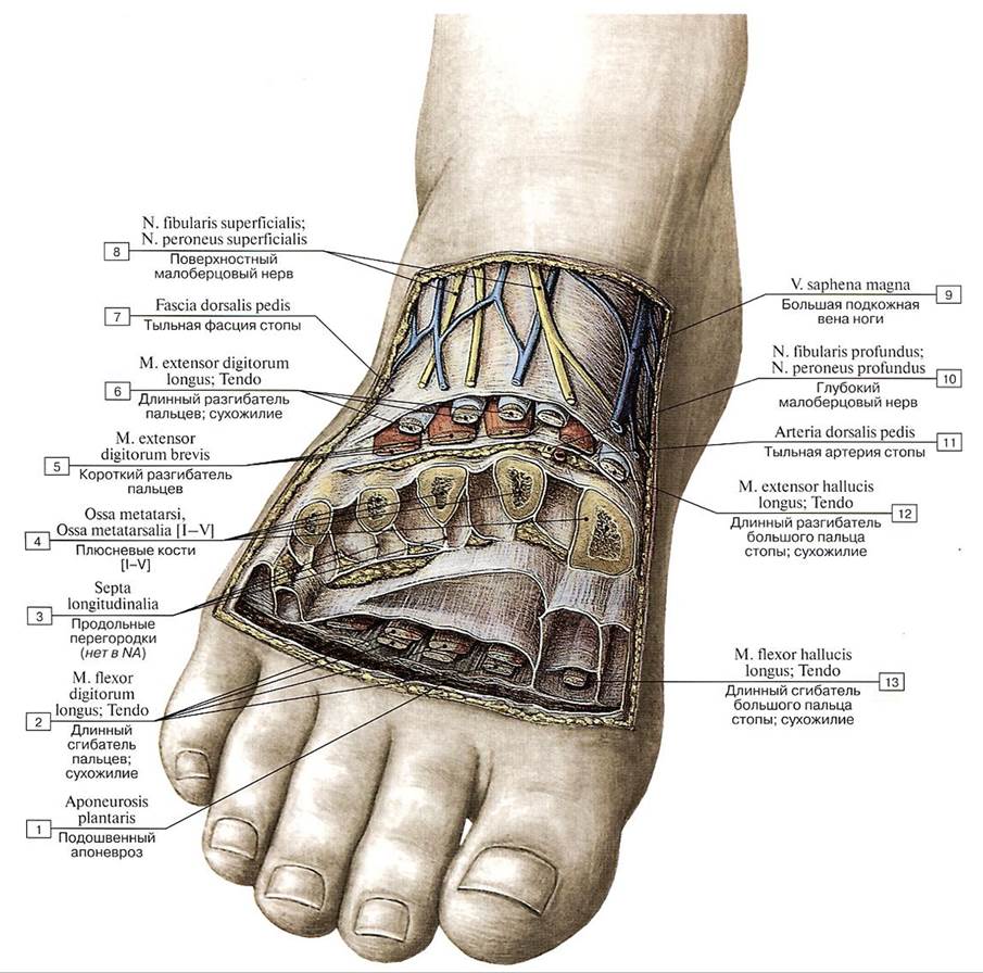 Фасциальные пространства стопы, правой, на поперечном распиле:
1 — Plantar aponeurosis; 2— Flexor digitorum longus; Tendon; 3— Longitudinal septa; 4— Metatarsals [I-V]; 5— Extensor digitorum brevis; 6 — Extensor digitorum longus; Tendon; 7 — Dorsal fascia of foot; 8— Superficial fibular nerve; Superficial peroneal nerve; 9 — Great saphenous vein; Long saphenous vein; 10 — Deep fibular nerve; Deep peroneal nerve; 11 — Dorsalis pedis artery; Dorsal artery of foot; 12— Extensor hallucis longus; Tendon; 13 — Flexor hallucis longus; Tendon
Фасциальные пространства стопы, правой, на поперечном распиле:
1 — Plantar aponeurosis; 2— Flexor digitorum longus; Tendon; 3— Longitudinal septa; 4— Metatarsals [I-V]; 5— Extensor digitorum brevis; 6 — Extensor digitorum longus; Tendon; 7 — Dorsal fascia of foot; 8— Superficial fibular nerve; Superficial peroneal nerve; 9 — Great saphenous vein; Long saphenous vein; 10 — Deep fibular nerve; Deep peroneal nerve; 11 — Dorsalis pedis artery; Dorsal artery of foot; 12— Extensor hallucis longus; Tendon; 13 — Flexor hallucis longus; Tendon
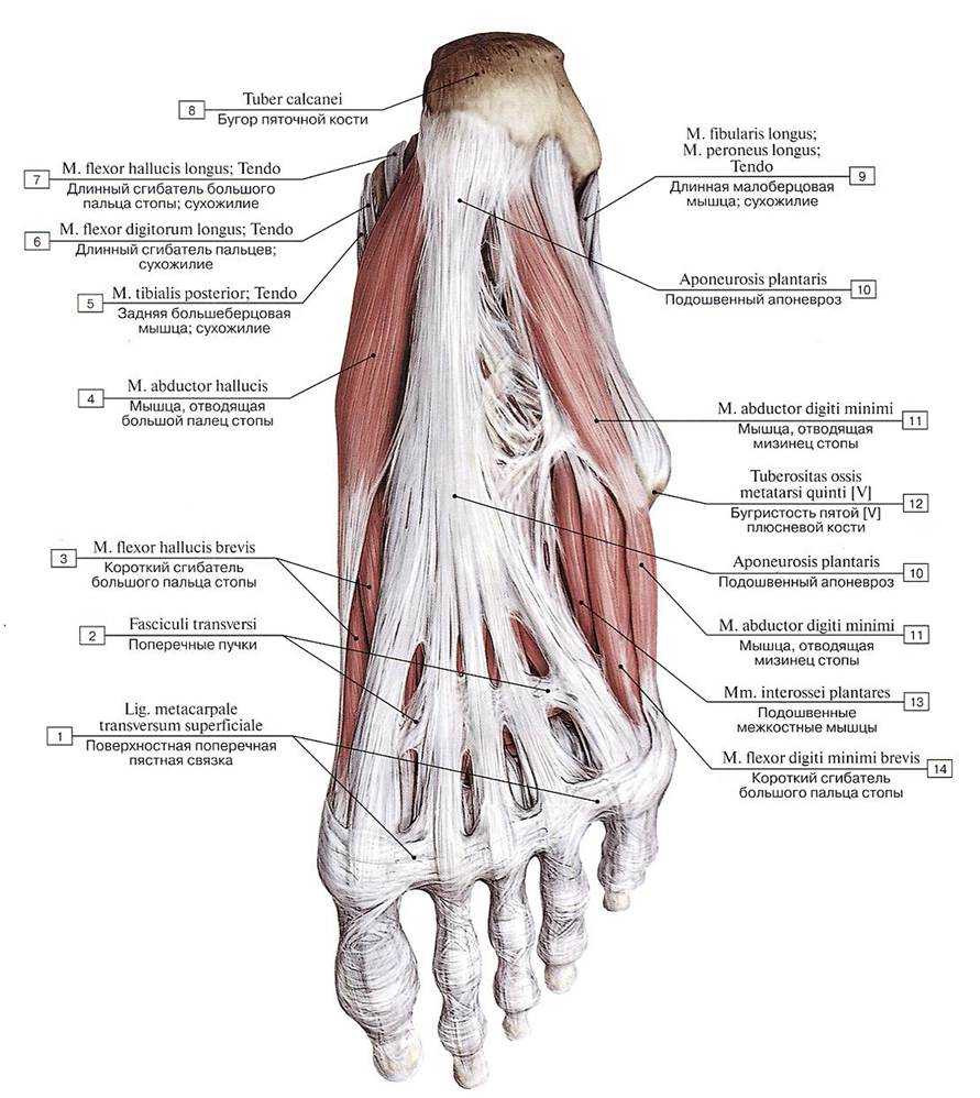 Подошвенный апоневроз и подошвенные мышцы стопы, правой, вид снизу:
1 — Superficial transverse metacarpal ligament; 2 — Transverse fascicles; 3 — Flexor hallucis brevis; 4 — Abductor hallucis; 5 — Tibialis posterior; Tendon; 6 — Flexor digitorum longus; Tendon; 7— Flexor hallucis longus; Tendon; 8— Calcaneal tuber; 9 — Fibularis longus; Peroneus longus; Tendon; 10 — Plantar aponeurosis; 11 — Abductor digiti minimi; 12—Tuberosity of fifth metatarsal bone [V]; 13 — Plantar interossei; 14 — Flexor digiti minimi brevis
Подошвенный апоневроз и подошвенные мышцы стопы, правой, вид снизу:
1 — Superficial transverse metacarpal ligament; 2 — Transverse fascicles; 3 — Flexor hallucis brevis; 4 — Abductor hallucis; 5 — Tibialis posterior; Tendon; 6 — Flexor digitorum longus; Tendon; 7— Flexor hallucis longus; Tendon; 8— Calcaneal tuber; 9 — Fibularis longus; Peroneus longus; Tendon; 10 — Plantar aponeurosis; 11 — Abductor digiti minimi; 12—Tuberosity of fifth metatarsal bone [V]; 13 — Plantar interossei; 14 — Flexor digiti minimi brevis
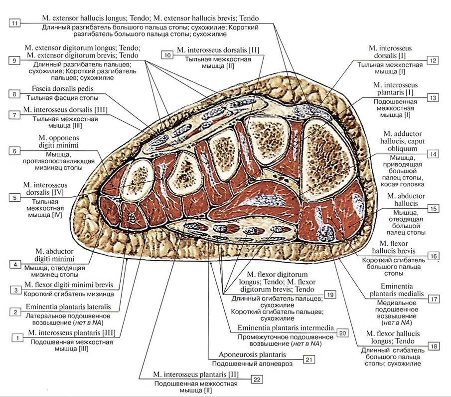 Поперечный распил стопы, левой, на уровне плюсны:
1 — Plantar interosseus [III]; 2 — Lateral plantar eminence; 3 — Flexor digiti minimi brevis; 4 — Abductor digiti minimi; 5 — Dorsal interosseus [IV]; 6 — Opponens digiti minimi; 7 — Dorsal interosseus [III]; 8 — Dorsal fascia of foot; 9 — Extensor digitorum longus; Tendon; Extensor digitorum brevis; Tendon; 10 — Dorsal interosseus [Il]; 11 — Extensor hallucis longus; Tendon; Extensor hallucis brevis; Tendon; 12 — Dorsal interosseus [I]; 13 — Plantar interosseus [I]; 14 — Adductor hallucis, oblique head; 15— Abductor hallucis; 16 — Flexor hallucis brevis; 17— Medial plantar eminence; 18— Flexor hallucis longus; Tendon; 19 — Flexor digitorum longus; Tendon; Flexor digitorum brevis; Tendon; 20 — Intermediate plantar eminence; 21 — Plantar aponeurosis; 22— Plantar interosseus [II]
Поперечный распил стопы, левой, на уровне плюсны:
1 — Plantar interosseus [III]; 2 — Lateral plantar eminence; 3 — Flexor digiti minimi brevis; 4 — Abductor digiti minimi; 5 — Dorsal interosseus [IV]; 6 — Opponens digiti minimi; 7 — Dorsal interosseus [III]; 8 — Dorsal fascia of foot; 9 — Extensor digitorum longus; Tendon; Extensor digitorum brevis; Tendon; 10 — Dorsal interosseus [Il]; 11 — Extensor hallucis longus; Tendon; Extensor hallucis brevis; Tendon; 12 — Dorsal interosseus [I]; 13 — Plantar interosseus [I]; 14 — Adductor hallucis, oblique head; 15— Abductor hallucis; 16 — Flexor hallucis brevis; 17— Medial plantar eminence; 18— Flexor hallucis longus; Tendon; 19 — Flexor digitorum longus; Tendon; Flexor digitorum brevis; Tendon; 20 — Intermediate plantar eminence; 21 — Plantar aponeurosis; 22— Plantar interosseus [II]
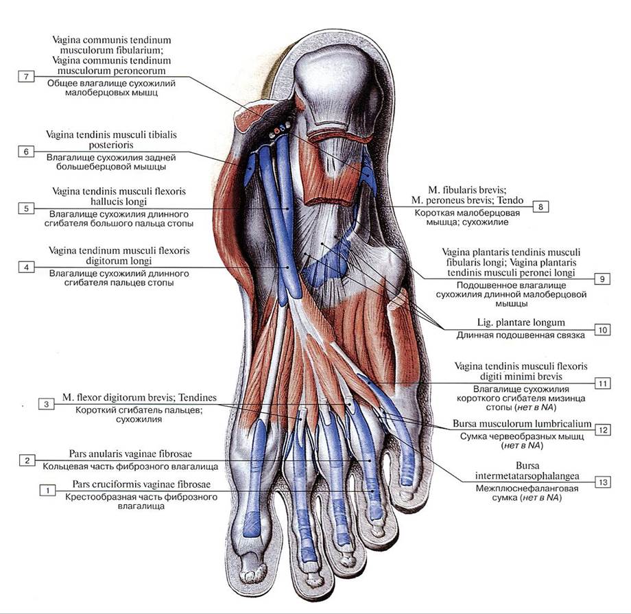 Синовиальные влагалища сухожилий мышц- сгибателей стопы, правой, вид снизу:
1 — Cruciform part of fibrous sheath; 2 — Anular part of fibrous sheath; 3 — Flexor digitorum brevis; Tendons; 4 —- Tendinous sheath of flexor digitorum longus; 5 — Tendinous sheath of flexor hallucis longus; 6 — Tendinous sheath of tibialis posterior; 7 — Common tendinous sheath of fibulares; Common tendinous sheath of peronei; 8 — Fibularis brevis; Peroneus brevis; Tendon; 9 — Plantar tendinous sheath of fibularis longus; Plantar tendinous sheath of peroneus longus; 10 — Long plantar ligament; 11 — Tendinous sheath of extensor digiti minimi brevis; 12 — Lumbrical bursa; 13 — Intermetatarsophalangeal bursa
Синовиальные влагалища сухожилий мышц- сгибателей стопы, правой, вид снизу:
1 — Cruciform part of fibrous sheath; 2 — Anular part of fibrous sheath; 3 — Flexor digitorum brevis; Tendons; 4 —- Tendinous sheath of flexor digitorum longus; 5 — Tendinous sheath of flexor hallucis longus; 6 — Tendinous sheath of tibialis posterior; 7 — Common tendinous sheath of fibulares; Common tendinous sheath of peronei; 8 — Fibularis brevis; Peroneus brevis; Tendon; 9 — Plantar tendinous sheath of fibularis longus; Plantar tendinous sheath of peroneus longus; 10 — Long plantar ligament; 11 — Tendinous sheath of extensor digiti minimi brevis; 12 — Lumbrical bursa; 13 — Intermetatarsophalangeal bursa
Страницы:
1 2 3 4 5 6 7 8 9 10 11 12 13 14 15 16 17 18